. 2009 May 29;284(22):14904-13.
doi: 10.1074/jbc.M809180200. Epub 2009 Mar 25.

Airway epithelial inflammation-induced endoplasmic reticulum Ca2+ store expansion is mediated by X-box binding protein-1

Affiliations

Airway epithelial inflammation-induced endoplasmic reticulum Ca2+ store expansion is mediated by X-box binding protein-1

Mary E B Martino et al. J Biol Chem. .

Abstract

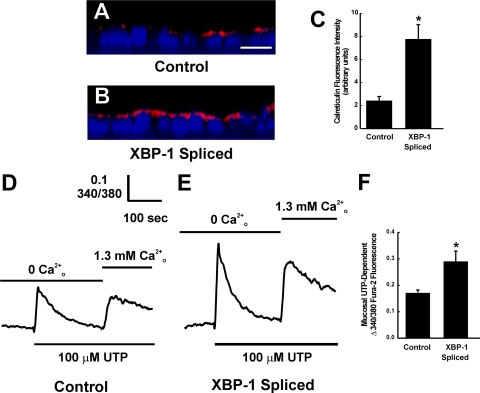
Inflamed cystic fibrosis (CF) human bronchial epithelia (HBE), or normal HBE exposed to supernatant from mucopurulent material (SMM) from CF airways, exhibit endoplasmic reticulum (ER)/Ca(2+) store expansion and amplified Ca(2+)-mediated inflammation. HBE inflammation triggers an unfolded protein response (UPR) coupled to mRNA splicing of X-box binding protein-1 (XBP-1). Because spliced XBP-1 (XBP-1s) promotes ER expansion in other cellular models, we hypothesized that XBP-1s is responsible for the ER/Ca(2+) store expansion in inflamed HBE. XBP-1s was increased in freshly isolated infected/inflamed CF in comparison with normal HBE. The link between airway epithelial inflammation, XBP-1s, and ER/Ca(2+) store expansion was then addressed in murine airways challenged with phosphate-buffered saline or Pseudomonas aeruginosa. P. aeruginosa-challenged mice exhibited airway epithelial ER/Ca(2+) store expansion, which correlated with airway inflammation. P. aeruginosa-induced airway inflammation triggered XBP-1s in ER stress-activated indicator (ERAI) mice. To evaluate the functional role of XBP-1s in airway inflammation linked to ER/Ca(2+) store expansion, control, XBP-1s, or dominant negative XBP-1 (DN-XBP-1) stably expressing 16HBE14o(-) cell lines were used. Studies with cells transfected with an unfolded protein response element (UPRE) luciferase reporter plasmid confirmed that the UPRE was activated or inhibited by expression of XBP-1s or DN-XBP-1, respectively. Expression of XBP-1s induced ER/Ca(2+) store expansion and potentiated bradykinin-increased interleukin (IL)-8 secretion, whereas expression of DN-XBP-1 inhibited bradykinin-dependent IL-8 secretion. In addition, expression of DN-XBP-1 blunted SMM-induced ER/Ca(2+) store expansion and SMM-induced IL-8 secretion. These findings suggest that, in inflamed HBE, XBP-1s is responsible for the ER/Ca(2+) store expansion that confers amplification of Ca(2+)-dependent inflammatory responses.


Figures

FIGURE 1.
FIGURE 1.
Native CF HBE exhibit increased XBP-1 mRNA splicing. A, Southern blot illustrating that XBP-1 mRNA splicing is increased in native infected/inflamed CF HBE as compared with non-infected/inflamed normal native HBE. Data are representative of three tissue codes from normal and CF HBE. B, compilation of the XBP-1 mRNA splicing data expressed as a percentage of XBP-1 mRNA splicing from normal epithelia.
FIGURE 2.
FIGURE 2.
Airway inflammation induced by P. aeruginosa infection promotes ER Ca2+ store expansion in murine airway epithelia in vivo. A and D, epithelia without airway inflammation from PBS-exposed wild type mice; B and E, epithelia from P. aeruginosa-infected mice in the presence of airway inflammation. C and F, epithelia from P. aeruginosa-infected mice in the absence of airway inflammation. A–C, green stain, calreticulin (ER Ca2+ store marker); blue stain,4′,6-diamidino-2-phenylindole-labeled nuclei. D–F, hematoxylin and eosin stain. Bar, 10 μm. P.a., P. aeruginosa.
FIGURE 3.
FIGURE 3.
Airway inflammation induced by P. aeruginosa infection promotes XBP-1 mRNA splicing in murine airway epithelia in vivo. Differential interference contrast (A, C, and E) and Venus expression (B, D, and F) in airway epithelia from non-inflamed, PBS-challenged (A and B), inflamed, P. aeruginosa (P.a.)-challenged (C and D) and non-inflamed, P.a.-challenged (E and F) ERAI mice airways. Arrows point to airway epithelia. Bar, 20 μm. G, compiled data for Venus fluorescence intensity, expressed per surface epithelial area, from all groups. *, p < 0.05, P.a.-challenged and inflamed versus PBS-challenged, non-inflamed.
FIGURE 4.
FIGURE 4.
SMM exposure induces IL-8 secretion coupled to XBP-1 mRNA splicing in polarized 16HBE14o- cultures. A, time course for mucosal PBS or SMM on serosal IL-8 secretion; *, p < 0.05, SMM versus PBS-treated cultures. B, Southern blot illustrating the time course for mucosal PBS or SMM on XBP-1 mRNA splicing.
FIGURE 5.
FIGURE 5.
Constitutive activation or inhibition of the UPRE in airway epithelia expressing XBP-1s or DN-XBP-1. A UPRE luciferase reporter was transfected into stable 16HBE14o- cell lines expressing control vector, XBP-1s, or DN-XBP-1. Cultures were exposed 48 h later to vehicle or 5 μg/ml tunicamycin for an additional 24 h. Luciferase activity, assessed by conventional methods, was normalized to CMV-LacZ. Data are expressed as fold-change of relative luciferase activity versus untreated empty vector-expressing cultures.
FIGURE 6.
FIGURE 6.
Expression of XBP-1s promotes morphological and functional expansion of ER Ca2+ stores in airway epithelia. A and B, XZ confocal images illustrating the immunofluorescent stain of calreticulin in polarized 16HBE14o- cultures expressing control vector or XBP-1s vector, respectively. Calreticulin is stained in red. Nuclei are stained in blue with 4′,6-diamidino-2-phenylindole. Bar, 10 μm. C, compiled data for calreticulin fluorescence from the two groups. D and E, representative tracings from 100 μm mucosal UTP-induced intracellular Ca2+ mobilization in polarized 16HBE14o- cultures loaded with fura-2 and expressing control vector or XBP-1s vector, respectively. First and second phases of Ca2+ mobilization in the absence or presence of extracellular Ca2+ illustrate ER Ca2+ release and capacitative calcium entry, respectively. F, compiled data for mucosal UTP-dependent ER Ca2+ release from the two groups.
FIGURE 7.
FIGURE 7.
Expression of DN-XBP-1 inhibits SMM-induced morphological and functional expansion of ER Ca2+ stores in airway epithelia. A, XZ confocal images illustrating the immunofluorescent stain of calreticulin in polarized 16HBE14o- cultures expressing control vector or DN-XBP-1 vector and exposed for 48 h to mucosal PBS or SMM. Calreticulin is stained in red. Nuclei are stained in blue with 4′,6-diamidino-2-phenylindole. Bar, 10 μm. B, compiled data for calreticulin fluorescence from the four groups. C–F, representative tracings from 100 μm mucosal UTP-induced intracellular Ca2+ mobilization in polarized 16HBE14o- cultures loaded with fura-2 and expressing control vector (C and D) or DN-XBP-1 vector (E and F). Cultures were exposed for 48 h to mucosal PBS (C and E) or SMM (D and F). First and second phases of Ca2+ mobilization in the absence or presence of extracellular Ca2+ illustrate ER Ca2+ release and capacitative calcium entry, respectively. G, compiled data for mucosal UTP-dependent ER Ca2+ release from the four groups.
FIGURE 8.
FIGURE 8.
Additional evidence that SMM-induced airway epithelial ER expansion is mediated by XBP-1s. A, representative Western blot of the ER marker PDI from polarized 16HBE14o- cultures expressing control, XBP-1s, or DN-XBP-1 vectors and exposed for 48 h to mucosal PBS or SMM. B, compiled data from the Western blot PDI signals from all groups.
FIGURE 9.
FIGURE 9.
XBP-1s constitutively promotes IL-8 secretion and plays a functional role in SMM- and BK-stimulated IL-8 secretion in airway epithelia. A, serosal IL-8 secretion from polarized 16HBE14o- cultures expressing control vector or DN-XBP-1 vector exposed for 24 h to mucosal PBS or SMM. *, p < 0.05 control vector + SMM versus control vector + PBS. B, serosal IL-8 levels from polarized 16HBE14o- cultures expressing control, XBP-1s, or DN-XBP-1 vectors resulting from 8 h exposure to vehicle or 5 μm mucosal BK. *, p < 0.05 XBP-1s versus control vector.

References

    1. Ribeiro, C. M. P., Paradiso, A. M., Carew, M. A., Shears, S. B., and Boucher, R. C. (2005) J. Biol. Chem. 280 10202-10209 - PubMed
    1. Ribeiro, C. M. P., Paradiso, A. M., Schwab, U., Perez-Vilar, J., Jones, L., O'Neal, W., and Boucher, R. C. (2005) J. Biol. Chem. 280 17798-17806 - PubMed
    1. Kaufman, R. J. (1999) Genes Dev. 13 1211-1233 - PubMed
    1. Mori, K. (2000) Cell 101 451-454 - PubMed
    1. Patil, C., and Walter, P. (2001) Curr. Opin. Cell Biol. 13 349-355 - PubMed

Publication types

MeSH terms