. 2009 May 22;284(21):13987-4000.
doi: 10.1074/jbc.M901758200. Epub 2009 Mar 25.

MDM2 acts downstream of p53 as an E3 ligase to promote FOXO ubiquitination and degradation

Affiliations

MDM2 acts downstream of p53 as an E3 ligase to promote FOXO ubiquitination and degradation

Wei Fu et al. J Biol Chem. .

Abstract

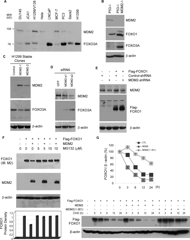
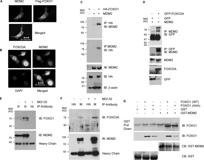
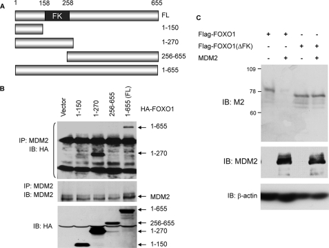
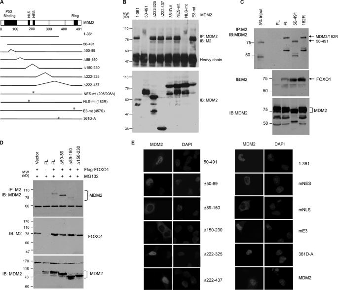
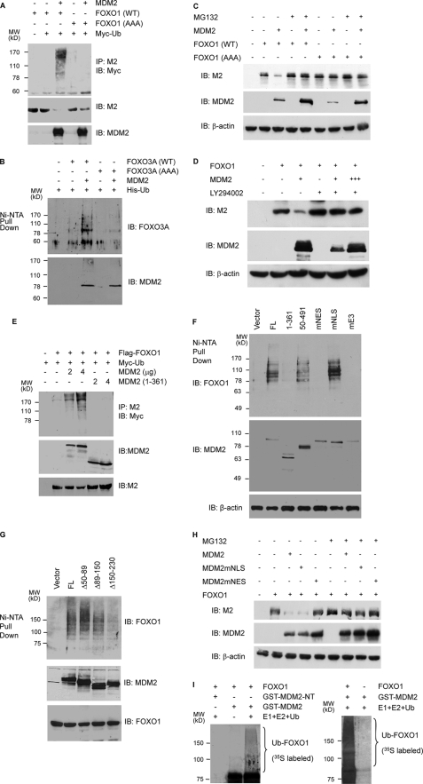
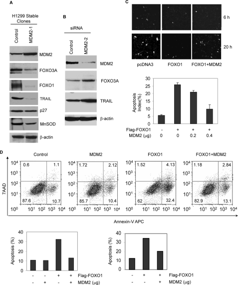
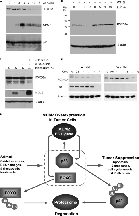
Members of the FOXO (forkhead O) class of transcription factors are tumor suppressors that also control aging and organismal life span. Mammalian FOXO degradation is proteasome-mediated, although the ubiquitin E3 ligase for FOXO factors remains to be defined. We show that MDM2 binds to FOXO1 and FOXO3A and promotes their ubiquitination and degradation, a process apparently dependent on FOXO phosphorylation at AKT sites and the E3 ligase activity of MDM2. Binding of MDM2 to FOXO occurs through the region of MDM2 that directs its cellular trafficking and the forkhead box of FOXO1. MDM2 promotes the ubiquitination of FOXO1 in a cell-free system, and its knockdown by small interfering RNA causes accumulation of endogenous FOXO3A protein in cells and enhances the expression of FOXO target genes. In cells stably expressing a temperature-sensitive p53 mutant, activation of p53 by shifting to permissive temperatures leads to MDM2 induction and degradation of endogenous FOXO3A. These data suggest that MDM2 acts as an ubiquitin E3 ligase, downstream of p53, to regulate the degradation of mammalian FOXO factors.


Figures

FIGURE 1.
FIGURE 1.
MDM2 promotes the degradation of FOXO in a proteasome-dependent manner. A, inverse correlation between MDM2 and FOXO3A protein expression in different cancer cell lines and MEFs. Cellular extracts were subjected to immunoblotting with anti-FOXO3A antibody (H-144). The membrane was washed and reprobed with anti-MDM2 (SMP-14). B, inverse correlation between MDM2 and FOXO in p53 null and p53 and MDM2 double null MEFs. Cellular extracts were subjected to immunoblotting with antibodies against FOXO1 (H-128), MDM2 (2A10), FOXO3A (H-144), and β-actin (AC-74), as indicated. C, stable expression of MDM2 decreases endogenous FOXO3A. Extracts of H1299 cells stably expressing MDM2 (MDM2-1 and MDM2-2) or empty vector (Control) were subjected to immunoblotting with the indicated antibodies. D, knockdown of endogenous MDM2 by siRNA increases the level of endogenous FOXO3A protein. H1299/V138 cells were transfected with 2 μg of siRNA against GFP or MDM2 (MDM2-s1 and MDM2-s2). Cellular extracts were prepared and subjected to immunoblotting with the indicated antibodies. E, knockdown of endogenous MDM2 by shRNA increases the level of ectopic FOXO1 protein. H1299/V138 cells were transfected with 0.5 μg of FLAG-FOXO1 together with 2 μg of shRNA vectors against MDM2 (MDM2-shRNA) or β-tubulin (Control-shRNA). Cellular extracts were prepared and subjected to immunoblotting with the indicated antibodies. F, ectopic MDM2 decreases the level of cotransfected FOXO1 protein, which is relieved by MG132. DU145 cells were transfected with 0.8 μg of FLAG-FOXO1 and 1.5 μg of MDM2. 24 h posttransfection, cells were incubated with or without MG132 for 6 h. Cellular extracts were subjected to immunoblotting (IB) with indicated antibodies. G, overexpression of MDM2 decreases the half-life of FOXO1 protein. H1299 cells were transfected with 0.8 μg of FLAG-FOXO1 and 1.5 μg of control vector, full-length (MDM2), or C-terminal truncated MDM2 (MDM2(1–361)). 24 h post-transfection, cells were treated with cycloheximide (CHX) for the indicated length of time. Cellular extracts containing equal amount of proteins were subjected to immunoblotting (lower panels). The immunoblotting signals were quantified by computer-based density scanning. The FOXO1 signals were normalized with cognate β-actin signals and plotted (graph).
FIGURE 2.
FIGURE 2.
MDM2 interacts with FOXO1 and FOXO3A in vivo and in vitro. A, colocalization of ectopic MDM2 and FLAG-FOXO1 in p53 and MDM2 double null MEFs. The fibroblasts were transfected with 0.5 μg of FLAG-FOXO1 and 0.5 μg of MDM2 and fixed. The colocalization was determined by immunofluorescence staining with anti-MDM2 and M2 anti-FLAG antibodies and visualized by deconvolution imaging. B, colocalization of endogenous MDM2 and FOXO3A. H1299 cells were fixed and subjected to immunofluorescence staining with DAPI, anti-MDM2, and anti-FOXO3A, as indicated. The colocalization was visualized by confocal imaging. C, interaction between ectopic FOXO1 and MDM2. H1299 cells were transfected with 2 μg of HA-FOXO1, 2 μg of MDM2, or both. 24 h posttransfection, cellular extracts were prepared and subjected to co-precipitations (IP) and immunoblotting (IB) with the indicated antibodies. D, interaction of ectopic FOXO3A and MDM2. H1299 cells were transfected with 2 μg of MDM2 and 2 μg of GFP or GFP-FOXO3A. 24 h posttransfection, cellular extracts were prepared and subjected to co-precipitations and immunoblotting with the indicated antibodies. E, interaction of endogenous MDM2 and FOXO1 in HI299/V138 cells. HI299/V138 cells were cultured at a permissive temperature (32 °C) overnight and treated with vehicle (DMSO) or MG132 for 6 h. Cellular extracts were subjected to co-immunoprecipitations, followed by immunoblotting with the indicated antibodies. M, anti-MDM2; HA, anti-HA (control). F, interaction of endogenous MDM2 and FOXO3A. HEK293T cells in a 100-mm dish were treated with DMSO or MG132 for 6 h. Then cellular extracts were prepared and subjected to co-immunoprecipitations, followed by immunoblotting with the indicated antibodies. M, anti-MDM2; HA, anti-HA (control). G, in vitro interaction between FOXO1 and MDM2. FOXO1 produced in in vitro transcription-coupled translation reactions was incubated with either GST or GST-MDM2 fusion proteins. Pull-down assays were performed with magnetic glutathione beads, followed by immunoblotting with anti-FOXO1 antibody. The amount of GST and GST-MDM2 proteins used in the pull-down assays was visualized by Coomassie Blue staining after SDS-PAGE (bottom).
FIGURE 3.
FIGURE 3.
The forkhead box of FOXO1 is required for MDM2 binding. A, diagram of the different HA-FOXO1 deletion constructs used in the interaction studies. FL, full-length FOXO1. B, co-immunoprecipitation (IP) analyses. H1299 cells were transfected with MDM2 and the indicated HA-FOXO1 vectors. Cellular extracts (lower panel) or anti-MDM2 immunoprecipitates (upper panels) were subjected to immunoblotting (IB) with the indicated antibodies. C, PC3 cells were transfected with 2 μg of FLAG-FOXO1 or FLAG-FOXO1 deleted of the forkhead box (FOXO1ΔFK) together with 2 μg of control vector or MDM2. Cellular extracts were subjected to immunoblotting with the indicated antibodies. The FOXO(ΔFK) expressed at a higher level than wild type, and reduced amounts of cellular extracts were used as indicated by lower level of β-actin.
FIGURE 4.
FIGURE 4.
The MDM2 sequence involved in the interaction with FOXO1 is defined to the region that contains nuclear localization and export sequences. A, a diagram showing different MDM2 mutant constructs used in the interaction studies. B, co-immunoprecipitations in the absence of proteasome inhibitors. H1299 cells were transfected with 2 μg of FLAG-FOXO1 and 2 μg of MDM2 mutants. Anti-MDM2 immunoprecipitates (upper panels) or cellular extracts (lower panel) were subjected to immunoblotting with the indicated antibodies. C and D, co-immunoprecipitation in the presence of MG132. H1299 cells were transfected with 2 μg of FLAG-FOXO1 and 2.5 μg of the MDM2 mutants and treated with MG132 for 6 h. Cellular extracts or anti-FLAG precipitates were subjected to immunoblotting with indicated antibodies. E, cellular localization of MDM2 mutants. H1299 cells were transfected with the indicated MDM2 constructs and immunostained with a MDM2 antibody. DAPI staining shows the nucleus.
FIGURE 5.
FIGURE 5.
MDM2 promotes the ubiquitination and degradation of FOXOs in a manner dependent on AKT-mediated phosphorylations and cytoplasmic localization. A, MDM2 promotes the ubiquitination of FOXO1 in vivo. H1299 cells were transfected with the indicated vectors. Cellular extracts were prepared, and FOXO1 ubiquitination was determined by precipitations (IP) with M2 anti-FLAG followed by immunoblotting (IB) with a Myc antibody (upper panel). The level of FOXO1 and MDM2 expression (lower panels) was determined by immunoblotting of the cellular extracts. B, MDM2 promotes the ubiquitination of FOXO3A in vivo. H1299 cells were transfected as indicated. Cellular extracts were subjected to Ni2+-NTA bead pull-down assays followed by immunoblotting with an HA antibody (upper panel). The level of MDM2 expression was determined by immunoblotting of the cellular extracts (lower panel). C, MDM2 promotes the FOXO1 degradation. H1299 cells were transfected with 2 μg of the indicated plasmids. 24 h later, the cells were treated with either DMSO (–) or 10 μm MG132 for 6 h. Cellular extracts were subjected to immunoblotting with the indicated antibodies. D, effect of LY294002 on the ability of MDM2 to decrease FOXO1 expression. PC3 cells were transfected with 2 μg of FLAG-FOXO1 and 2 (+) and 6 (+++) μg of MDM2. 15 h later, the cells were treated with either DMSO or 10 μm LY294002 for 8 h. Cellular extracts were subjected to immunoblotting with the indicated antibodies. E, dose-dependent effect of MDM2 on FOXO1 ubiquitination and the requirement of its carboxyl-terminal RING domain. p53 and MDM2 double null MEFs were transfected with the indicated vectors, and the ubiquitination of FOXO1 and the level of FOXO1 and MDM2 expression were determined by immunoblotting of the cellular extracts. F and G, ubiquitination of FOXO1 requires MDM2 E3 ligase activity, cytoplasmic localization, and interaction with FOXO1. p53 and MDM2 double null MEFs were transfected with the indicated vectors, and the ubiquitination of FOXO1 as well as the level of FOXO1 and MDM2 expression were determined by immunoblotting of the cellular extracts. H, the dependence on cytoplasmic localization for the ability of MDM2 to decrease FOXO1 level. H1299 cells were transfected with 2 μg of FLAG-FOXO1 and MDM2. 24 h later, the cells were treated with either DMSO or 10 μm MG132 for 6 h. I, MDM2 promotes FOXO1 polyubiquitination in vitro. GST-MDM2 and GST-MDM2-(1–150) (GST-NT) were produced in E. coli and bound to glutathione-agarose beads, and the ubiquitination of the bound FOXO1 protein was assayed as described under “Materials and Methods.” Ubiquitinated FOXO1 was visualized by autoradiography as a high molecular weight smear above the unmodified FOXO1 band.
FIGURE 6.
FIGURE 6.
MDM2 suppresses the expression of FOXO1 target gene expression and protects cells from FOXO1 induced apoptosis. A, stable expression of MDM2 suppresses the expression of FOXO target genes. Whole cell extracts of H1299 cells and MDM2 stable clones were subjected to immunoblotting with the indicated antibodies. B, MDM2 siRNA enhances the induction of TRAIL by FOXO1. H1299 cells were transfected with siRNA against GFP or MDM2. 48 h posttransfection, cellular extracts were prepared and subjected to immunoblotting with the indicated antibodies. C, MDM2 protects cells from FOXO1-induced apoptosis, as measured by manual counting. H1299 cells were transfected with 0.2 μg of pLNCE and 0.5 μg of FLAG-FOXO1 together with or without 1 μg of MDM2. Then the cells were fixed and stained with DAPI. Representative micrographs were captured by a CCD camera attached to a fluorescence microscope (upper panels). The apoptotic index of GFP-positive cells was determined by scoring 300 GFP-positive cells for chromatin condensation and nuclear fragmentation. Triplicate samples were analyzed per data point, and the graph represents three independent experiments. D, MDM2 protects cells from FOXO1-induced apoptosis, as measured by flow cytometry. H1299 cells were transfected with GFP-spectrin and FLAG-FOXO1 with or without MDM2. Transfected (GFP-positive) cells were separated from nontransfected (GFP-negative) cells by FACS-based sorting. The apoptotic index of GFP-positive cells was determined with the Annexin-V kit. The flow cytometry profile of a representative experiment was shown. Data from two independent analyses are shown as bar graphs. MnSOD, manganese superoxide dismutase.
FIGURE 7.
FIGURE 7.
p53 induces the degradation of FOXO3A protein through MDM2. A, p53 expression causes the down-regulation of endogenous FOXO3A protein. H1299/V138 cells at restrictive temperature were shifted to 32 °C for the indicated length of time. Cellular extracts were prepared and subjected to immunoblotting. B, MG132 blocks the protein expression decrease of FOXO3A. H1299/V138 cells were shifted to 32 °C as in A but treated with MG132 for 6 h before cellular extracts were prepared for immunoblotting. C, knockdown of MDM2 partially relieves p53-induced FOXO3A down-regulation. H1299/V138 cells were transfected with scrambled or MDM2 siRNA. 24 h posttransfections, cells were shifted to 32 °C and cultured for 18 h. Then immunoblotting was performed with the indicated antibodies. D, effect of p53 on FOXO3a protein stability after UV treatment. Wild-type (WT) and p53 null MEFs were treated with UV (0.15 J) and then with 10 μg/ml cycloheximide (CHX). The cells were harvested at the indicated time points. Cellular extracts were subjected to immunoblotting for the indicated proteins. E, a working model describing how cellular DNA damage response suppresses tumorigenesis through the functions of p53 and FOXO proteins and how MDM2 overexpression in cancer cells promotes tumor growth by inducing the degradation of both p53 and FOXO proteins through the ubiquitin-proteasome pathway. The model also describes the possibility that after DNA repair is accomplished, MDM2 induced by p53 can turn off both p53 and FOXO factors as a feedback mechanism.

References

    1. Katoh, M., and Katoh, M. (2004) Int. J. Oncol. 25 1495–1500 - PubMed
    1. Medema, R. H., Kops, G. J., Bos, J. L., and Burgering, B. M. (2000) Nature 404 782–787 - PubMed
    1. Nakamura, N., Ramaswamy, S., Vazquez, F., Signoretti, S., Loda, M., and Sellers, W. R. (2000) Mol. Cell. Biol. 20 8969–8982 - PMC - PubMed
    1. Schmidt, M., Fernandez de Mattos, S., van der Horst, A., Klompmaker, R., Kops, G. J., Lam, E. W., Burgering, B. M., and Medema, R. H. (2002) Mol. Cell. Biol. 22 7842–7852 - PMC - PubMed
    1. Modur, V., Nagarajan, R., Evers, B. M., and Milbrandt, J. (2002) J. Biol. Chem. 277 47928–47937 - PubMed

MeSH terms

Substances