. 2009 May 22;284(21):14126-35.
doi: 10.1074/jbc.M900687200. Epub 2009 Mar 25.

Coordinated activation of the origin licensing factor CDC6 and CDK2 in resting human fibroblasts expressing SV40 small T antigen and cyclin E

Affiliations

Coordinated activation of the origin licensing factor CDC6 and CDK2 in resting human fibroblasts expressing SV40 small T antigen and cyclin E

Elena Sotillo et al. J Biol Chem. .

Abstract

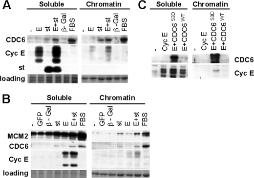
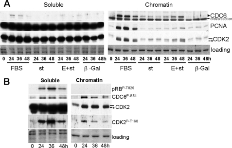
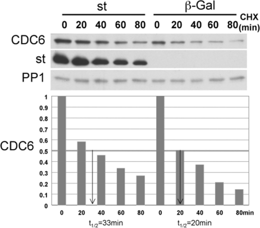
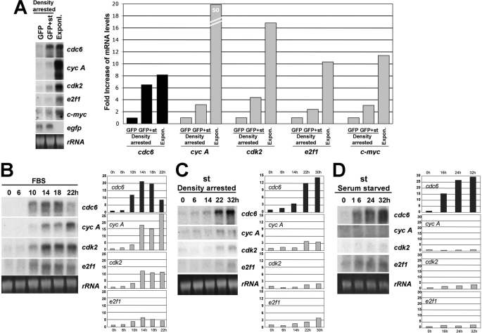
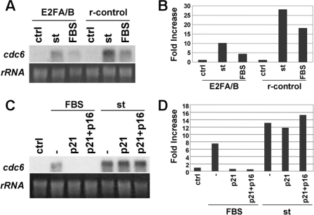
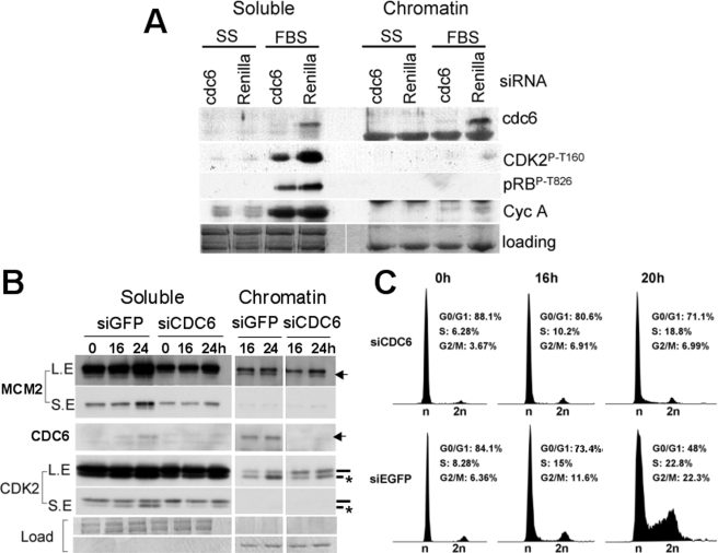
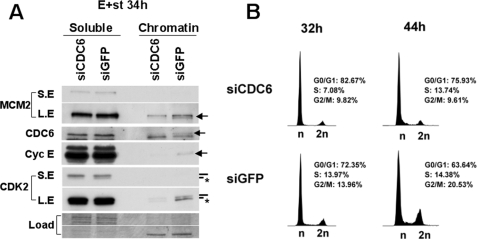
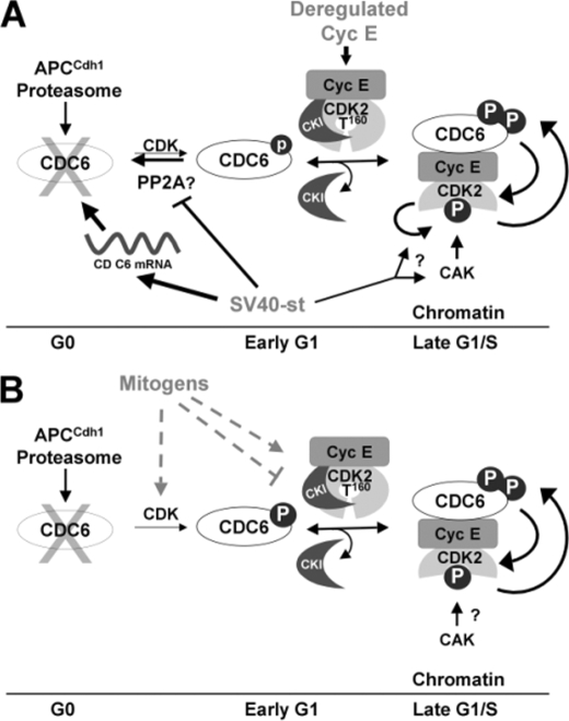
We have previously shown that SV40 small t antigen (st) cooperates with deregulated cyclin E to activate CDK2 and bypass quiescence in normal human fibroblasts (NHF). Here we show that st expression in serum-starved and density-arrested NHF specifically induces up-regulation and loading of CDC6 onto chromatin. Coexpression of cyclin E results in further accumulation of CDC6 onto chromatin concomitantly with phosphorylation of CDK2 on Thr-160 and CDC6 on Ser-54. Investigation of the mechanism leading to CDC6 accumulation and chromatin loading indicates that st is a potent inducer of cdc6 mRNA expression and increases CDC6 protein stability. We also show that CDC6 expression in quiescent NHF efficiently promotes cyclin E loading onto chromatin, but it is not sufficient to activate CDK2. Moreover, we show that CDC6 expression is linked to phosphorylation of the activating T loop of CDK2 in serum-starved NHF stimulated with mitogens or ectopically expressing cyclin E and st. Our data suggest a model where the combination of st and deregulated cyclin E result in cooperative and coordinated activation of both an essential origin licensing factor, CDC6, and an activity required for origin firing, CDK2, resulting in progression from quiescence to S phase.


Figures

FIGURE 1.
FIGURE 1.
st induces expression and loading of CDC6 onto chromatin in quiescent NHF, and CDC6 facilitates loading of cyclin E. BJ-hTERT fibroblasts were serum-starved for 72 h (A) or grown to high density as described under “Experimental Procedures” (B and C) and then transduced with the indicated adenoviruses or re-stimulated with FBS. Forty-eight hours following transduction, cells were harvested, and chromatin-bound and soluble fractions were obtained as described under “Experimental Procedures.” 15 μg of the soluble protein fractions and the corresponding equivalent chromatin-bound fractions were analyzed by Western blot using the indicated antibodies. Same exposure times are shown for both fractions. Coomassie Blue staining of the gel is shown as a loading control. Relevant proteins are indicated. Note that as reported earlier exogenously expressed cyclin E migrates as a series of bands that migrate faster that endogenous cyclin E (12).
FIGURE 2.
FIGURE 2.
st expression in serum-starved NHF induces CDC6 expression and loading, which is enhanced by coexpression of cyclin E. The kinetics of phosphorylation of CDK2 on Thr-160 and CDK2 substrates is distinct in the soluble and chromatin-bound fraction. BJ-hTERT fibroblasts were serum-starved for 72 h and transduced with indicated adenoviruses (A) or re-stimulated with serum (A and B) and harvested at indicated time points. Soluble and chromatin-bound proteins were obtained and simultaneously analyzed by Western blot. Same exposure times are shown for both fractions. Coomassie Blue staining is shown as a loading control. Relevant proteins are indicated. The CDK2 band corresponding to phosphorylated Thr-160 is indicated with an asterisk.
FIGURE 3.
FIGURE 3.
st increases CDC6 protein stability. Density-arrested BJ-hTERT fibroblasts stably expressing Myc-tagged CDC6 from a retroviral plasmid were transduced with adenovirus expressing st or control β-galactosidase. Forty hours after transduction, cells were treated with 10 μm CHX and harvested at the indicated time points. Whole cell lysates were analyzed by Western blot. CDC6 signal was quantified relative to time 0 and normalized to the PP1 signal, which is stable through the time course.
FIGURE 4.
FIGURE 4.
cdc6 mRNA expression is potently up-regulated by st in serum-starved and density-arrested BJ-hTERT cells. A, BJ-hTERT cells were grown to high density and transduced with 40 MOI of adenovirus expressing st and/or control EGFP and harvested 30 h later. Exponentially growing cells were used as controls. Levels of expression of indicated mRNAs were analyzed by Northern blot as described under “Experimental Procedures.” Radioactive signals were measured by densitometry, quantified with NIH Image J, and represented as fold increase. B, BJ-hTERT fibroblasts were synchronized by serum starvation for 72 h, re-stimulated with 10% FBS and harvested at indicated time points for Northern blot analysis as in A. C, BJ-hTERT cells were grown to high density, transduced with adenovirus expressing st and harvested at indicated time points after transduction and treated as in A. D, BJ-hTERT cells were serum-starved for 72 h, transduced with adenovirus expressing st and harvested at indicated time points after transduction and treated as in A.
FIGURE 5.
FIGURE 5.
st induces CDC6 mRNA expression in a CDK-independent, E2F-dependent manner. A, BJ-hTERT cells stably expressing a dominant negative E2F/pRB mutant chimera or the empty expression vector (r-control) (see text) were serum-starved for 72 h immediately after antibiotic selection and re-stimulated with FBS or transduced with control or st adenovirus. Cells were harvested for RNA analysis 40 h later. B, radioactive signals were quantified, normalized by ribosomal RNA loading, and represented as fold increase in mRNA as in Fig. 4. C, BJ-hTERT cells were serum-starved for 48 h and transduced with adenovirus expressing p21 and/or p16. Twenty-four hours later, cells were re-stimulated with FBS or transduced with st adenovirus. Forty hours later, cells were harvested for Northern blot analysis. D, radioactive signal was quantified as in B.
FIGURE 6.
FIGURE 6.
CDC6 expression is coupled to phosphorylation of CDK2 on Thr-160 and exit from quiescence induced by serum. A, BJ-hTERT cells were transfected with siRNAs targeting CDC6 or renilla luciferase in conditions of serum starvation. Forty-eight hours later, cells were restimulated with 10% FBS, and 48 h after restimulation, cells were harvested and lysed for soluble/chromatin-bound protein fractionation and Western blot analysis. B and C, BJ-hTERT cells were transfected with siRNAs targeting CDC6 or EGFP in conditions of serum starvation. Seventy-two hours later, cells were re-stimulated with FBS and harvested at the indicated time points. Cell pellets were divided in two for soluble/chromatin-bound protein fractionation and Western blot analysis (B) and PI/FACS analysis (C). A and B, same exposure times are shown for both fractions. B, CDK2 band corresponding to phosphorylated Thr-160 is indicated with an asterisk. LE indicates long exposure. SE indicates short exposure. C, percent of cells in each cell cycle phase is indicated.
FIGURE 7.
FIGURE 7.
CDC6 expression is required for phosphorylation of CDK2 on Thr-160 and exit from quiescence induced by deregulated cyclin E and st co-expression. BJ-hTERT cells were transfected with the indicated siRNA and serum-starved as in Fig. 6. Subsequently the cells were transduced with adenovirus expressing cyclin E and st and harvested 34 h later. Cell pellets were divided in two for soluble/chromatin-bound protein fractionation and Western blot analysis (A) and PI/FACS analysis (B). A, same exposure times are shown for both fractions. Relevant proteins are indicated. The CDK2 band corresponding to phosphorylated Thr-160 is indicated with an asterisk. LE indicates long exposure. SE indicates short exposure. B, percent of cells in each cell cycle phase is indicated.
FIGURE 8.
FIGURE 8.
Model of events linking CDC6 and CDK2 activation during exit from quiescence induced by cyclin E and st coexpression or mitogens. A, expression of st and deregulated cyclin E expression in quiescent NHF result in cooperative activation of the essential origin licensing factor CDC6 and coordinated CDK2 activation. st potently induces CDC6 mRNA expression and to a lower extent CDC6 phosphorylation on a CDK site(s) and stabilization. st also facilitates loading of CKI free cyclin E/CDK2 onto chromatin, which correlates with CDK2 phosphorylation on the activating T loop. CDC6 may facilitate CAK access to the CDK2 T loop, which could be prevented by CKIs. Alternatively st may induce CDK2 autophosphorylation on Thr-160 (arrow with question mark; see text). Coexpression of cyclin E and st also enhances CDC6 phosphorylation and loading onto chromatin suggesting positive feedback loops leading to activation of CDK2 and CDC6 accumulation (indicated by reciprocal arrows). B, mitogens may use similar mechanisms to activate CDK2 on chromatin.

References

    1. Malumbres, M., and Barbacid, M. (2005) Trends Biochem. Sci. 30 630–641 - PubMed
    1. Graña, X., Garriga, J., and Mayol, X. (1998) Oncogene 17 3365–3383 - PubMed
    1. Mailand, N., and Diffley, J. F. (2005) Cell 122 915–926 - PubMed
    1. Geng, Y., Yu, Q., Sicinska, E., Das, M., Schneider, J. E., Bhattacharya, S., Rideout, W. M., Bronson, R. T., Gardner, H., and Sicinski, P. (2003) Cell 114 431–443 - PubMed
    1. Geng, Y., Lee, Y. M., Welcker, M., Swanger, J., Zagozdzon, A., Winer, J. D., Roberts, J. M., Kaldis, P., Clurman, B. E., and Sicinski, P. (2007) Mol. Cell 25 127–139 - PubMed

Publication types

MeSH terms

LinkOut - more resources