. 2009 Jun;37(10):3243-53.
doi: 10.1093/nar/gkp190. Epub 2009 Mar 24.

Evolutionary changes in the Leishmania eIF4F complex involve variations in the eIF4E-eIF4G interactions

Affiliations

Evolutionary changes in the Leishmania eIF4F complex involve variations in the eIF4E-eIF4G interactions

Yael Yoffe et al. Nucleic Acids Res. 2009 Jun.

Abstract

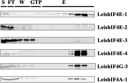
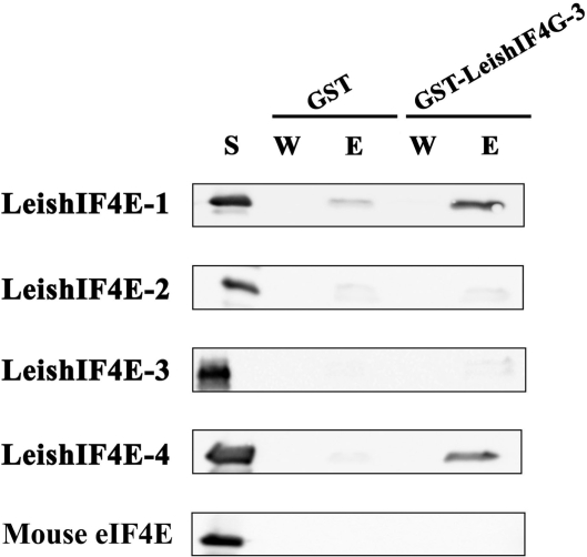
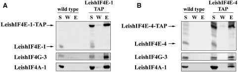
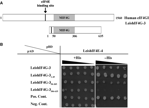
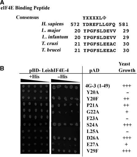
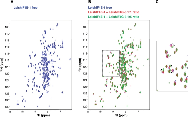
Translation initiation in eukaryotes is mediated by assembly of the eIF4F complex over the m(7)GTP cap structure at the 5'-end of mRNAs. This requires an interaction between eIF4E and eIF4G, two eIF4F subunits. The Leishmania orthologs of eIF4E are structurally diverged from their higher eukaryote counterparts, since they have evolved to bind the unique trypanosomatid cap-4 structure. Here, we characterize a key eIF4G candidate from Leishmania parasites (LeishIF4G-3) that contains a conserved MIF4G domain. LeishIF4G-3 was found to coelute with the parasite eIF4F subunits from an m(7)GTP-Sepharose column and to bind directly to LeishIF4E. In higher eukaryotes the eIF4E-eIF4G interaction is based on a conserved peptide signature [Y(X(4))Lphi], where X is any amino acid and Phi is a hydrophobic residue. A parallel eIF4E-binding peptide was identified in LeishIF4G-3 (20-YPGFSLDE-27). However, the binding motif varies extensively: in addition to Y20 and L25, binding strictly requires the presence of F23, whereas the hydrophobic amino acid (Phi) is dispensable. The LeishIF4E-LeishIF4G-3 interaction was also confirmed by nuclear magnetic resonance (NMR) studies. In view of these diversities, the characterization of the parasite eIF4E-eIF4G interaction may not only serve as a novel target for inhibiting Leishmaniasis but also provide important insight for future drug discovery.


Figures

Figure 1.
Figure 1.
Leishmania major cap-binding complex purification. Leishmania major cells were lyzed and the soluble protein extract (S) was loaded over an m7GTP-Sepharose column. After collection of the flowthrough (FT) the column was washed (W) with CB (see Materials and Methods section) and with CB containing 100 μM GTP (GTP). The proteins were eluted (E) with CB containing 500 mM NaCl. Equal samples from the different fractions were separated by SDS–PAGE and analyzed by western blot using specific antibodies against the different subunits of the Leishmania cap-binding complex (LeishIF4E-1 through LeishIF4E-4, LeishIF4G-3, LeishIF4A-1). Similar results were obtained when the m7GTP-Sepharose column was eluted with free m7GTP.
Figure 2.
Figure 2.
LeishIF4G-3 comigrates with LeishIF4E-1 and LeishIF4E-4 by sucrose gradients analysis. Extracts of L. major cells grown at 26°C, and after exposure to 35°C for 18 h were loaded on 10–50% sucrose gradients. (A) Pattern of polysome migration on sucrose gradients. Fractions were collected from the top of the gradient and the O.D260nm was monitored. (26°C, continuous line; 35°C, broken line). Proteins were recovered by TCA precipitation, and immunoblotted with antibodies raised against LeishIF4E-1, LeishIF4E-4, LeishIF4G-3 as well as with anti-rpS6, for detection of fractions that contain complexes containing 40S particles. Western analysis was done on fractions of cells that were grown at 26°C (B) and 35°C (C).
Figure 3.
Figure 3.
LeishIF4G-3 interacts with LeishIF4E-1 and LeishIF4E-4 in vitro. GST or GST-LeishIF4G-3 fusion protein were immobilized on Glutathione-Agarose beads. The beads were incubated with supernatant of bacteria that express the different LeishIF4E isoforms or the mouse eIF4E ortholog (S). After extensive washes (W) the protein complexes were eluted (E), and analyzed by western blot with specific antibodies against the different eIF4Es.
Figure 4.
Figure 4.
LeishIF4G-3-FLAG pulls down LeishIF4E-4. Leishmania major cells were transfected with pX-LeishIF4G-3-FLAG and cell lines expressing the FLAG tagged LeishIF4G-3 were selected in the presence of 200 μg/ml G418. The cells were lyzed by sonication and the soluble fractions (S) were incubated with anti-FLAG beads. After incubation for 3 h, the beads were washed (W), the proteins were eluted (E), separated by SDS–PAGE and analyzed by western blot.
Figure 5.
Figure 5.
LeishIF4G-3 and LeishIF4A-1 copurifiy with TAP-tagged LeishIF4E-1 and LeishIF4E-4. Leishmania major wild-type or transgenic cells expressing TAP-tagged LeishIF4E-1 (A) and LeishIF4E-4 (B) were lyzed and the soluble supernatant (S) was loaded on streptavidin-Sepharose beads. The beads were washed, eluted with biotin and repurified in tandem over IgG-Sepharose beads (LeishIF4E-1) or over m7GTP-Sepharose beads (LeishIF4E-4). The corresponding beads were washed (W) and eluted (E, see Materials and Methods section). The W and E lanes were loaded with 20% of the wash and eluate volumes, respectively. All proteins were fractionated by SDS–PAGE (12%) and subjected to western blot analysis using antibodies specific against LeishIF4E-1, LeishIF4E-4, LeishIF4G-3 and LeishIF4A-1.
Figure 6.
Figure 6.
Monitoring the LeishIF4G-3 interaction with LeishIF4E-4 by the yeast two-hybrid assay. (A) Scheme of LeishIF4G-3 highlighting the location of its eIF4E-binding region and the MIF4G domain, as compared with the human eIF4GI. (B) Yeast cells were cotransformed with pBD-LeishIF4E-4 and pAD fused to different fragments of LeishIF4G-3 as indicated (1–49, 50–305, 306–635), or with positive or negative control plasmids (see Material and Methods section for details). The cells were cultured in Liquid SD-2 (-Trp/-Leu/+His), and 3-fold dilutions were spotted on SD-2 or SD-3 (-Trp/-Leu/-His) containing 2 mM 3-AT; the plates were incubated at 30°C for 10 days.
Figure 7.
Figure 7.
Effect of mutations in the LeishIF4G-3 consensus peptide on binding to LeishIF4E-4. (A) Sequence conservation of the consensus peptide [Y(X4)LΦ] of LeishIF4G-3 in different trypanosomatids (L. major, L. infantum, T. cruzi, T. brucei), as compared with the human peptide (H. sapiens). (B) Yeast cells were cotransformed with pBD-LeishIF4E-4 and pAD fused to the LeishIF4G-31–49 fragment carrying different point mutations in the consensus peptide. The cells were cultured as described in the legend of Figure 6, and growth in the absence of histidine served as an indication for binding between LeishIF4G-3 and LeishIF4E-4.
Figure 8.
Figure 8.
Binding of a LeishIF4G-3 peptide to LeishIF4E-1. (A) 15N, 1HTROSY-HSQC spectra of 15N-labeled LeishIF4E-1 (250 µM) in the presence of m7GTP (5 mM) for stabilization (LeishIF4E-1 free, in blue). (B) Overlays of 15N, 1HTROSY-HSQC spectra of 15N-LeishIF4E-1 free with increasing amounts of non-labeled LeishIF4G-3 peptide (NYLEPPYPGFSLDEVVRR). The concentrations of the LeishIF4G-3 added were 0 µM (blue, ratio 1:0), 250 µM (red, ratio 1:1) and 1.25 mM (green, ratio 1:5). (C) Enlargement of a region displaying the chemical shift differences between the spectra that is boxed in (B) in dashed lines. Peaks movements are indicated by arrows.

References

    1. Shapira M, McEwen JG, Jaffe CL. Temperature effects on molecular processes which lead to stage differentiation in Leishmania. EMBO J. 1988;7:2895–2901. - PMC - PubMed
    1. Zilberstein D, Shapira M. The role of pH and temperature on the development of Leishmania parasites. Annu. Rev. Microbiol. 1994;48:449–470. - PubMed
    1. Bates P. Axenic culture of Leishmania amastigotes. Parasitol. Today. 1993;9:143–146. - PubMed
    1. Barak E, Amin-Spector S, Gerliak E, Goyard S, Holland N, Zilberstein D. Differentiation of Leishmania donovani in host-free system: analysis of signal perception and response. Mol. Biochem. Parasitol. 2005;141:99–108. - PubMed
    1. Liang XH, Haritan A, Uliel S, Michaeli S. Trans- and cis-splicing in trypanosomatids: mechanism, factors, and regulation. Eukaryot. Cell. 2003;2:830–840. - PMC - PubMed

Publication types

MeSH terms