. 2009 Jun;83(12):6161-70.
doi: 10.1128/JVI.02488-08. Epub 2009 Mar 25.

Fas-mediated apoptotic signaling in the mouse brain following reovirus infection

Affiliations

Fas-mediated apoptotic signaling in the mouse brain following reovirus infection

Penny Clarke et al. J Virol. 2009 Jun.

Abstract

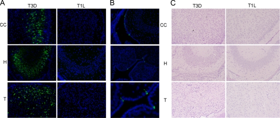
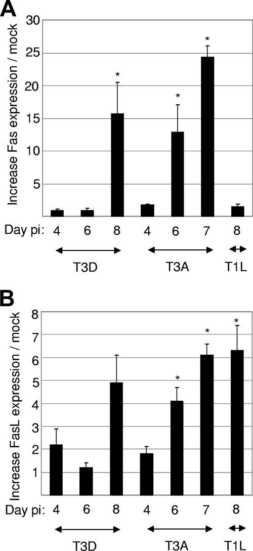
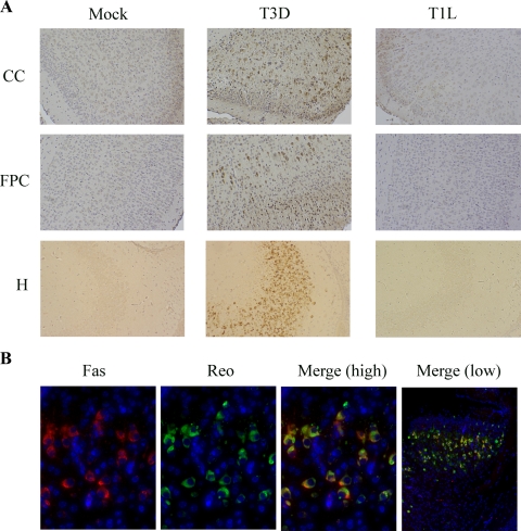
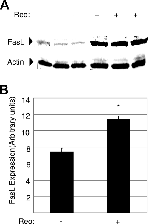
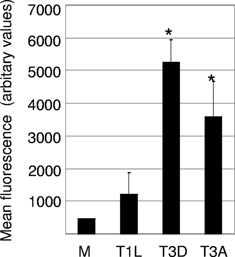
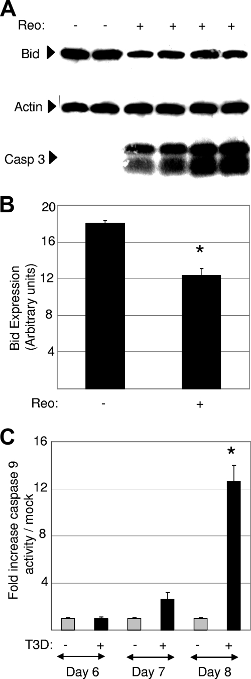
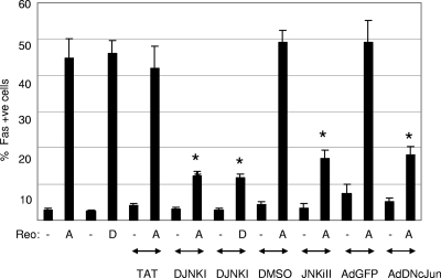
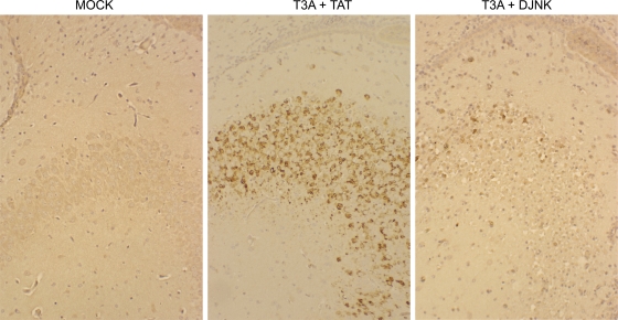
Type 3 (T3) reovirus strains induce apoptotic neuronal cell death and lethal encephalitis in infected mice. T3 strain Dearing (T3D)-induced apoptosis in primary neuronal cultures occurs by a Fas-mediated mechanism and requires the activation of caspase 8. We now show that Fas mRNA is upregulated in the brains of mice infected with encephalitic reovirus T3D and T3 strain Abney (T3A) but not following infection with nonencephalitic reovirus type 1 strain Lang. Fas is upregulated in regions of the brain that are injured during infection with T3 reovirus strains and colocalizes with virus antigen in individual neurons. In contrast, levels of FasL mRNA induced by encephalitic and nonencephalitic reovirus strains do not differ significantly. Caspase 8, the initiator caspase associated with Fas-mediated apoptosis, is activated in the cortex and hippocampal regions of both T3D- and T3A-infected mice. Furthermore, Bid cleavage and the activation of caspase 9 in the brains of T3D-infected mice suggest that the caspase 8-dependent activation of mitochondrial apoptotic signaling contributes to virus-induced apoptosis. We have previously shown that the inhibition of c-Jun N-terminal kinase (JNK) signaling blocks T3D-induced apoptosis and improves the outcome of virus-induced encephalitis. We now show that the reovirus-induced upregulation of Fas requires JNK signaling, thereby providing a link between reovirus-induced death receptor signaling and mitogen-activated protein kinase pathways and a potential mechanism for the therapeutic action of JNK inhibition.


Figures

FIG. 1.
FIG. 1.
Following intracerebral infection with T3D, virus antigen colocalizes with tissue injury in the cortex, hippocampus, and thalamus. (A) Immunohistochemical analysis indicates that reovirus antigen (green staining) is present in the cingulate cortex (CC), hippocampus (H), and thalamus (T) 8 days following intracerebral injection of T3D (103 PFU). In contrast, virus antigen is not expressed in these regions 8 days following intracerebral injection of T1L (103 PFU). (B) Viral antigen following T1L infection is found in the ependymal cells surrounding the lateral (top) and third (middle and bottom) ventricles. Tissue sections in A and B were incubated with rabbit polyclonal antireovirus antibody followed by FITC-labeled goat anti-rabbit antibody. Magnifications, ×400 (A and B, top and middle) and ×1,000 (B, bottom). The figure is representative of slides generated from at least three individual T3D- or T1L-infected animals. (C) Staining with hematoxylin and eosin indicates that tissue injury (neuronal cell death and minor inflammation) is present in the hippocampus (H), cingulate cortex (CC), and thalamus (T) 8 days following intracerebral injection of T3D (103 PFU). In contrast, injury is not evident in these areas 8 days following the intracerebral injection of T1L (103 PFU). Magnification, ×200. The figure is representative of slides generated from at least three individual T3D- or T1L-infected animals.
FIG. 2.
FIG. 2.
The level of Fas mRNA is upregulated in the brains of T3 reovirus-infected mice. mRNA was prepared from mouse brains following intracerebral injection of 103 PFU of encephalitic reovirus strains T3D (mRNA prepared on days 4, 6, and 8 postinfection [pi]) and T3A (mRNA prepared on days 4, 6, and 7 postinfection) and nonencephalitic reovirus strain T1L (mRNA prepared on day 8 postinfection). RT PCR was performed using primers designed to amplify Fas and FasL sequences. Cyclophilin was used as a control gene. The graphs show the mean increases in the levels of expression of Fas (A) or FasL (B) in reovirus-infected mouse brains compared to those seen in mock-infected brains. Error bars represent standard errors of the means. A minimum of three mock-infected and three reovirus-infected animals were used for each condition. Conditions generating statistically significant differences (P < 0.05) between reovirus-infected and mock-infected animals are indicated (*).
FIG. 3.
FIG. 3.
Fas is upregulated in the brains of T3D-infected mice in areas targeted by virus infection and colocalizes in individual cells with virus antigen. (A) Immunochemical analysis indicates that Fas (brown staining) is expressed in the cingulate cortex (CC), frontoparietal cortex (FPC), and hippocampus (H) 8 days following intracerebral injection of T3D reovirus (103 PFU). In contrast, Fas is not expressed in these regions following infection with T1L or in mock-infected mouse brains. Magnification, ×200. (B) Immunohistochemical analysis of tissue sections from the cingulate cortex demonstrates that Fas (red staining) and reovirus antigen (reo) (green staining) colocalize in individual cells (see yellow staining in merged image). Tissue sections were incubated with rabbit polyclonal antireovirus (reo) or mouse monoclonal anti-Fas primary antibody followed by FITC-labeled goat anti-rabbit or Cy3-labeled goat anti-mouse secondary antibody. Magnifications, ×400 (Fas, antireovirus, and merge [high]) and ×100 (merge [low]). The figure is representative of tissue sections from at least three individual T3D-, T1L-, and mock-infected animals.
FIG. 4.
FIG. 4.
The level of FasL is increased in T3A-infected mouse brains. Protein lysates were prepared from mouse brains 7 days following intracerebral injection of 103 PFU of encephalitic reovirus strain T3A. (A) Lysates were analyzed by Western blot analysis using antibody directed against FasL. Antibody directed against actin was used as a protein loading control. Each lane represents an individual mock-infected (−) or T3A-infected (+) animal. (B) Densitometric analysis was used to determine the level of FasL expression in T3A- and mock-infected mouse brains, with values normalized to actin levels. The graph shows the mean level of FasL expression in mock-infected compared to T3A-infected mouse brains. Error bars represent standard errors of the means. Conditions generating statistically significant differences (P < 0.05) between reovirus-infected and mock-infected animals are indicated (*).
FIG. 5.
FIG. 5.
The level of caspase 8 activity is increased in the brains of T3 reovirus-infected mice. Protein extracts were prepared from reovirus-infected and mock-infected (M) mice and were analyzed for caspase 8 activity using a fluorogenic substrate assay. The graph shows the mean fluorescence (caspase 8 activity). Error bars represent standard errors of the means. A minimum of four animals were used for each condition. Conditions generating statistically significant differences (P < 0.05) between reovirus-infected and mock-infected animals are indicated (*).
FIG. 6.
FIG. 6.
Reovirus infection of mouse brains results in the cleavage of Bid and the activation of caspase 9. (A) Protein lysates were prepared from mouse brains 8 days following intracerebral injection of 103 PFU of encephalitic reovirus strain T3D. Lysates were analyzed by Western blot analysis using antibody directed against Bid. Antibody directed against activated caspase 3 was used to demonstrate the presence of apoptosis, and antibody directed against actin was used as a protein loading control. Each lane represents an individual mock-infected (−) or T3D-infected (+) animal. (B) Densitometric analysis was used to compare Bid expression levels in T3D- and mock-infected mouse brains, with values normalized to actin levels. The graph shows the mean levels of Bid in mock-infected compared to T3D-infected mouse brains. Error bars represent standard errors of the means. Conditions generating statistically significant differences (P < 0.05) between reovirus-infected and mock-infected animals are indicated (*). (C) Protein extracts from T3D reovirus-infected mouse brains were also analyzed for caspase 9 activity. The graph shows the mean increase in caspase 9 activity levels in T3D-infected mouse brains compared to that of mock-infected controls. A minimum of three mock-infected and three reovirus-infected animals were used for each condition. Error bars represent standard errors of the means. Conditions generating statistically significant differences (P < 0.05) between reovirus-infected and mock-infected animals are indicated (*).
FIG. 7.
FIG. 7.
Reovirus-induced upregulation of Fas requires JNK signaling. HeLa cells were infected with encephalitic reovirus strains T3A and T3D (MOI of 20) in the presence of JNK inhibitor D-JNK-1 and JNKiII or an adenoviral vector expressing DN c-Jun (AdDNcJun). The percentage of cells expressing Fas was compared to that of reovirus-infected HeLa cells treated with control vehicles for D-JNKI-1 (a vector expressing TAT protein only) and JNKiII (dimethyl sulfoxide [DMSO]) or an adenoviral vector expressing GFP (AdGFP). After 30 h, cells were fixed and incubated with antibodies directed against Fas. The graph shows the mean percentages of immunoreactive cells from three independent experiments. Error bars represent standard errors of the means. Conditions generating statistically significant differences (P < 0.05) between treated and nontreated reovirus-infected cells are indicated (*).
FIG. 8.
FIG. 8.
JNK is required for Fas upregulation during reovirus encephalitis. Immunohistochemical analysis indicates that the treatment of mice with D-JNKI-1 (DJNK) (11 mg/kg/day for 10 days) blocks the reovirus (103 PFU)-induced expression of Fas (brown staining) seen in the hippocampus of animals treated with a control peptide (TAT) (11 mg/kg/day for 10 days). Fas is also not expressed in mock-infected mouse brains. The figure is representative of data from three individual T3D-infected mice treated with D-JNKI-1, three individual T3D-infected mice treated with a control peptide, and three individual mock-infected animals.

References

    1. Ashkenazi, A., and V. M. Dixit. 1998. Death receptors: signaling and modulation. Science 2811305-1308. - PubMed
    1. Baloul, L., S. Camelo, and M. Lafon. 2004. Up-regulation of Fas ligand (FasL) in the central nervous system: a mechanism of immune evasion by rabies virus. J. Neurovirol. 10372-382. - PubMed
    1. Beckham, J. D., R. J. Goody, P. Clarke, C. Bonny, and K. L. Tyler. 2007. Novel strategy for treatment of viral central nervous system infection by using a cell-permeating inhibitor of c-Jun N-terminal kinase. J. Virol. 816984-6992. - PMC - PubMed
    1. Brozovic, S., R. Sahoo, S. Barve, H. Shiba, S. Uriarte, R. S. Blumberg, and D. F. Kinane. 2006. Porphyromonas gingivalis enhances FasL expression via up-regulation of NFkappaB-mediated gene transcription and induces apoptotic cell death in human gingival epithelial cells. Microbiology 152797-806. - PubMed
    1. Brunner, T., R. J. Mogil, D. LaFace, N. J. Yoo, A. Mahboubi, F. Echeverri, S. J. Martin, W. R. Force, D. H. Lynch, C. F. Ware, et al. 1995. Cell-autonomous Fas (CD95)/Fas-ligand interaction mediates activation-induced apoptosis in T-cell hybridomas. Nature 373441-444. - PubMed

Publication types

MeSH terms