Clinical Trial
. 2009 Jun;83(11):5881-9.
doi: 10.1128/JVI.02345-08. Epub 2009 Mar 25.

Comparison of human and rhesus macaque T-cell responses elicited by boosting with NYVAC encoding human immunodeficiency virus type 1 clade C immunogens

Affiliations
Clinical Trial

Comparison of human and rhesus macaque T-cell responses elicited by boosting with NYVAC encoding human immunodeficiency virus type 1 clade C immunogens

Petra Mooij et al. J Virol. 2009 Jun.

Abstract

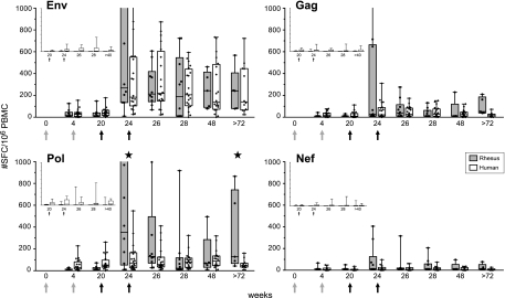
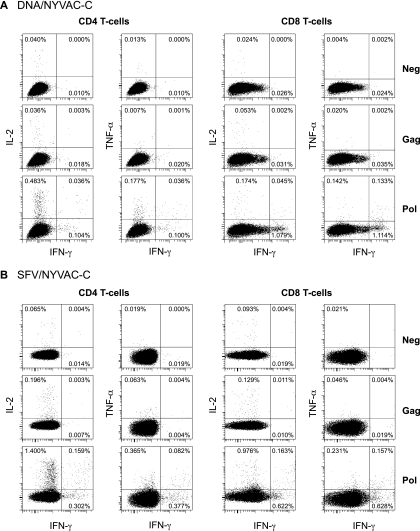
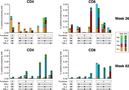
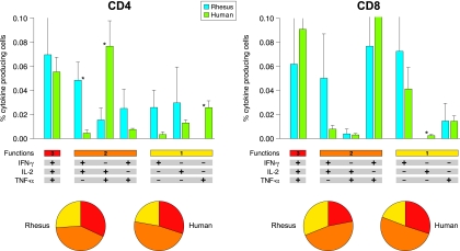
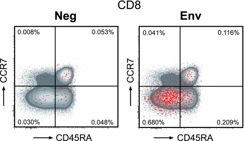
Rhesus macaques (Macaca mulatta) have played a valuable role in the development of human immunodeficiency virus (HIV) vaccine candidates prior to human clinical trials. However, changes and/or improvements in immunogen quality in the good manufacturing practice (GMP) process or changes in adjuvants, schedule, route, dose, or readouts have compromised the direct comparison of T-cell responses between species. Here we report a comparative study in which T-cell responses from humans and macaques to HIV type 1 antigens (Gag, Pol, Nef, and Env) were induced by the same vaccine batches prepared under GMP and administered according to the same schedules in the absence and presence of priming. Priming with DNA (humans and macaques) or alphavirus (macaques) and boosting with NYVAC induced robust and broad antigen-specific responses, with highly similar Env-specific gamma interferon (IFN-gamma) enzyme-linked immunospot assay responses in rhesus monkeys and human volunteers. Persistent cytokine responses of antigen-specific CD4(+) and CD8(+) T cells of the central memory as well as the effector memory phenotype, capable of simultaneously eliciting multiple cytokines (IFN-gamma, interleukin 2, and tumor necrosis factor alpha), were induced. Responses were highly similar in humans and primates, confirming earlier data indicating that priming is essential for inducing robust NYVAC-boosted IFN-gamma T-cell responses. While significant similarities were observed in Env-specific responses in both species, differences were also observed with respect to responses to other HIV antigens. Future studies with other vaccines using identical lots, immunization schedules, and readouts will establish a broader data set of species similarities and differences with which increased confidence in predicting human responses may be achieved.


Figures

FIG. 1.
FIG. 1.
Direct comparison of vaccine-induced antigen-specific IFN-γ ELISpot assay responses in rhesus monkeys and human volunteers. IFN-γ secretion by PBMC of individual DNA/NYVAC-immunized animals (n = 10) and human volunteers (n = 20) to HIV-1 clade C Env (two peptide pools), Gag (two peptide pools), Pol (three peptide pools), and Nef (one peptide pool) overlapping peptides was measured by ELISpot assay. Immunizations were given at weeks 0 and 4 (DNA; gray arrows) and weeks 20 and 24 (NYVAC; black arrows). Box-whisker plots show the interquartile ranges and medians (horizontal lines) of the groups at each time point. The last time point is week 82 for rhesus monkeys and week 72 for humans. Background responses (mean number of SFC plus 2 SD of triplicate assays with medium alone) were subtracted. Statistically significant differences (P < 0.05; Wilcoxon's rank sum test) between humans and rhesus monkeys are indicated by stars. (Insets) Antigen-specific IFN-γ responses of rhesus monkeys (first bars) and human volunteers (second bars) that were not primed with DNA but only immunized at week 20 and 24 (black arrows) with NYVAC expressing HIV-1 clade C Env, Gag, Pol, and Nef. The last time point is week 40 for rhesus monkeys and week 48 for humans. Values on the y axes range from 0 to 1,000 SFC/106 PBMC.
FIG. 2.
FIG. 2.
Vaccine-induced antigen-specific T-cell responses in time. IFN-γ, IL-2, and IL-4 secretion by PBMC of individual animals (dots) to HIV-1 clade C Env (two peptide pools), Gag (two peptide pools), Pol (three peptide pools), and Nef (one peptide pool) overlapping peptides was measured by ELISpot assay. Immunizations were given at weeks 0 and 4 (DNA or SFV priming) and weeks 20 and 24 (NYVAC boosting). Box-whisker plots show the interquartile ranges and medians (horizontal lines) of the groups at each time point. Background responses (mean number of SFC plus 2 SD of triplicate assays with medium alone) were subtracted. Responses of animals that were not primed (monitored from weeks 20 to 40), primed with DNA, or primed with SFV are presented in one graph for direct comparison. Statistically significant differences (P < 0.05; Wilcoxon's rank sum test) between the two priming groups are indicated by stars. (DNA priming > SFV priming) and diamonds (SFV priming > DNA priming).
FIG. 3.
FIG. 3.
Vaccine-induced antigen-specific intracellular cytokine production as measured by FACS analysis at week 82. Flow cytometry profiles of Gag- and Pol-specific CD4+ (left) and CD8+ (right) T cells able to secrete IFN-γ, IL-2, and TNF-α of a representative DNA-primed animal (A) and an SFV-primed animal (B) are shown.
FIG. 4.
FIG. 4.
Functional profiles of vaccine-induced antigen-specific CD4+ and CD8+ T cells. The results shown are from five animals of each group (DNA/NYVAC-C and SFV/NYVAC-C) at week 26 and week 82. Responses are grouped on the basis of the number of functions (producing one, two, or three cytokines simultaneously). All the possible combinations of cytokine responses are shown on the x axis. The responses to vaccine antigens (Env, Gag, Pol, and Nef) are color coded. Bars correspond to the mean number of different functionally distinct antigen-specific CD4+ or CD8+ T cells per group. Background responses have been subtracted.
FIG. 5.
FIG. 5.
Direct comparison of rhesus and human functional profiles of vaccine-induced Env-specific CD4+ and CD8+ T cells. The results shown were generated from five animals and eight human volunteers immunized with DNA/NYVAC-C at the peak of the response. Responses are grouped on the basis of the number of functions (producing one, two, or three cytokines simultaneously). All the possible combinations of cytokine responses are shown on the x axis. Bars correspond to the mean number of different functionally distinct Env-specific CD4+ or CD8+ T cells per group. Standard errors of the means are also presented. Each slice of the pie charts corresponds to the fraction of CD4+ or CD8+ T cells with a given number of functions (as shown below the graphs) within the total CD4+ and CD8+ T-cell populations. Statistically significant differences between rhesus macaques and human are indicated by asterisks (Mann-Whitney). Background responses have been subtracted.
FIG. 6.
FIG. 6.
Phenotypic analysis of vaccine-induced CD8+ T cells. The memory phenotype of antigen-specific T cells was determined in 10 animals (5 animals from each primed group). PBMC were unstimulated (Neg) or stimulated with Env peptides for 16 h and stained with IFN-γ, IL-2, TNF-α, CD3, CD4, CD8, CCR7, and CD45RA antibodies. A flow cytometry scatter plot of CD8+ T cells from animal Ri508 (DNA primed) at week 26 (peak) is shown. The red dots indicate the cytokine-producing vaccine-induced CD8+ T cells. Env-specific CD8+ T cells are of the EM (CD45RA CCR7) and EFF (CD45RA+ CCR7) phenotypes.

References

    1. Amara, R. R., C. Ibegbu, F. Villinger, D. C. Montefiori, S. Sharma, P. Nigam, Y. Xu, H. M. McClure, and H. L. Robinson. 2005. Studies using a viral challenge and CD8 T cell depletions on the roles of cellular and humoral immunity in the control of an SHIV-89.6P challenge in DNA/MVA-vaccinated macaques. Virology 343246-255. - PubMed
    1. Amara, R. R., F. Villinger, J. D. Altman, S. L. Lydy, S. P. O'Neil, S. I. Staprans, D. C. Montefiori, Y. Xu, J. G. Herndon, L. S. Wyatt, M. A. Candido, N. L. Kozyr, P. L. Earl, J. M. Smith, H. L. Ma, B. D. Grimm, M. L. Hulsey, J. Miller, H. M. McClure, J. M. McNicholl, B. Moss, and H. L. Robinson. 2001. Control of a mucosal challenge and prevention of AIDS by a multiprotein DNA/MVA vaccine. Science 29269-74. - PubMed
    1. Balla-Jhagjhoorsingh, S. S., G. Koopman, P. Mooij, W. Koornstra, S. McCormack, J. Weber, G. Pantaleo, and J. L. Heeney. 2004. Long-term persistence of HIV-1 vaccine-induced CD4+CD45RA−CD62L−CCR7− memory T-helper cells. AIDS 18837-848. - PubMed
    1. Bart, P. A., R. Goodall, T. Barber, A. Harari, A. Guimaraes-Walker, M. Khonkarly, N. C. Sheppard, Y. Bangala, M. J. Frachette, R. Wagner, P. Liljestrom, J. P. Kraehenbuhl, M. Girard, J. Goudsmit, M. Esteban, J. Heeney, Q. Sattentau, S. McCormack, A. Babiker, G. Pantaleo, and J. Weber. 2008. EV01: A phase I trial in healthy HIV negative volunteers to evaluate a clade C HIV vaccine, NYVAC-C undertaken by the EuroVacc Consortium. Vaccine 263153-3161. - PubMed
    1. Betts, M. R., M. C. Nason, S. M. West, S. C. De Rosa, S. A. Migueles, J. Abraham, M. M. Lederman, J. M. Benito, P. A. Goepfert, M. Connors, M. Roederer, and R. A. Koup. 2006. HIV nonprogressors preferentially maintain highly functional HIV-specific CD8+ T cells. Blood 1074781-4789. - PMC - PubMed

Publication types