. 2009 May;20(10):2563-71.
doi: 10.1091/mbc.e08-10-1019. Epub 2009 Mar 25.

Trf1 is not required for proliferation or functional telomere maintenance in chicken DT40 cells

Affiliations

Trf1 is not required for proliferation or functional telomere maintenance in chicken DT40 cells

Carol Cooley et al. Mol Biol Cell. 2009 May.

Abstract

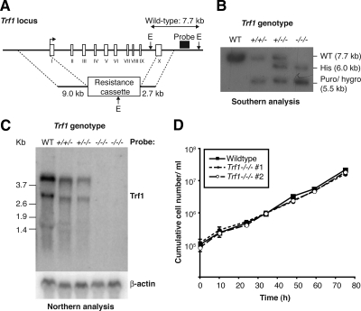
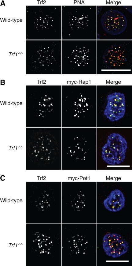
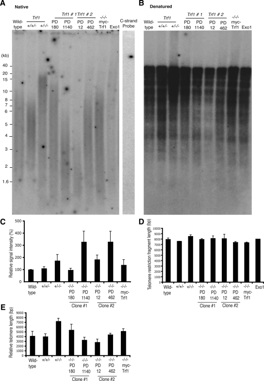
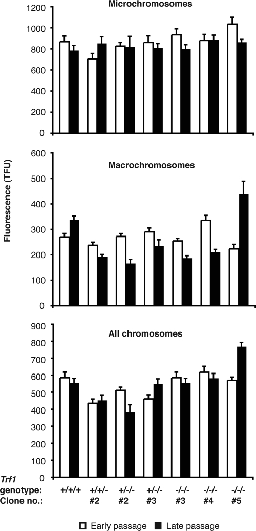
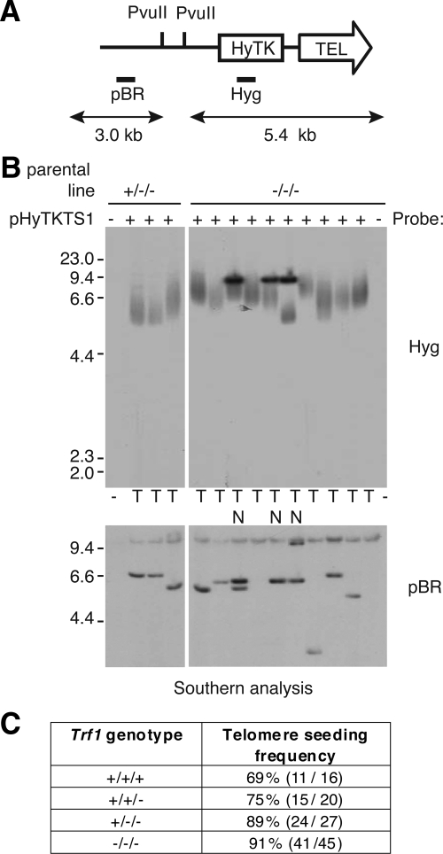
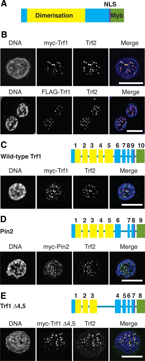
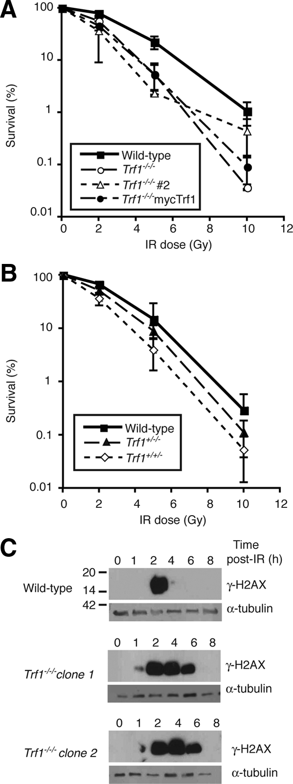
The telomere end-protection complex prevents the ends of linear eukaryotic chromosomes from degradation or inappropriate DNA repair. The homodimeric double-stranded DNA-binding protein, Trf1, is a component of this complex and is essential for mouse embryonic development. To define the requirement for Trf1 in somatic cells, we deleted Trf1 in chicken DT40 cells by gene targeting. Trf1-deficient cells proliferated as rapidly as control cells and showed telomeric localization of Trf2, Rap1, and Pot1. Telomeric G-strand overhang lengths were increased in late-passage Trf1-deficient cells, although telomere lengths were unaffected by Trf1 deficiency, as determined by denaturing Southern and quantitative FISH analysis. Although we observed some clonal variation in terminal telomere fragment lengths, this did not correlate with cellular Trf1 levels. Trf1 was not required for telomere seeding, indicating that de novo telomere formation can proceed without Trf1. The Pin2 isoform and a novel exon 4, 5-deleted isoform localized to telomeres in Trf1-deficient cells. Trf1-deficient cells were sensitive to DNA damage induced by ionizing radiation. Our data demonstrate that chicken DT40 B cells do not require Trf1 for functional telomere structure and suggest that Trf1 may have additional, nontelomeric roles involved in maintaining genome stability.


Figures

Figure 1.
Figure 1.
Gene targeting of Trf1 and preliminary phenotypic analysis. (A) Diagrammatic representation of the chicken Trf1 locus and gene targeting strategy. Exons are shown by white boxes and labeled with Roman numerals. E, relevant EcoRI sites. (B) Southern blot analysis of sequential Trf1 targeting steps in clones of the indicated genotypes. Expected sizes of the wild-type and targeted alleles are indicated at right. (C) Northern blot analysis of clones of the indicated genotypes hybridized with the full-length cDNA probes shown at right. Size markers at left are in kb. (D) Proliferation analysis of wild-type and two Trf1−/−/− clones. Data points are the mean ± SD of three separate experiments.
Figure 2.
Figure 2.
Microscopy analysis of shelterin components in Trf1-deficient cells. (A) Cells of the indicated genotype were fixed and subjected to in situ hybridization with a PNA anti-telomere probe (red) then stained with antibodies to Trf2 (green) and counterstained with DAPI (blue). Cells of the indicated genotype were transiently transfected with expression vectors for myc-Rap1 (B) or myc-Pot1 (C) and then fixed and stained with antibodies to myc (red) and to Trf2 (green) and counterstained with DAPI (blue). Scale bars, 10 μm.
Figure 3.
Figure 3.
Telomere maintenance and telomerase activity in Trf1-deficient cells. Autoradiogram showing native (A) and denatured (B) in-gel hybridizations of (TA2C3)4 G-strand probe to MspI-, HinfI-, and HaeIII-digested genomic DNA from the indicated cell lines at the population doublings (PD) indicated. As controls, wild-type samples were treated with ExoI or mung bean nuclease (MBN) before restriction digestion. Wild-type DNA was probed with a C-strand probe, which has sequence complementary to the G-strand probe. Trf1−/−/− myc-Trf1–expressing cells were passaged for ∼800–900 PD. (C) Histogram showing relative G-overhang signals. The signal in each lane was quantified with a phosphoimager and normalized against the signal detected in a standardized region in the denatured gel. Data represent the mean values + SEM of three independent experiments. (D) Histogram showing average telomere length for the indicated clones, as determined by analysis of the denaturing gel. Signals were measured using Image Quant, with each lane being divided into 40 boxes, and the weighted mean telomere length was calculated using the formula previously described (Wei et al., 2002). Data shown are from three independent experiments. (E) Histogram showing average telomere length for the indicated clones, as determined by analysis of the nondenaturing gel using the same approach as in D.
Figure 4.
Figure 4.
Q-FISH analysis of (TTAGGG)n fluorescence of micro- and macro-chromosomes in wild-type DT40 and Trf1 mutant cell lines. A Cy3-conjugated PNA probe complementary to the (TTAGGG)n repeat array was hybridized to DAPI-stained metaphase spreads of DT40 cells of the indicated genotypes. Fluorescence intensities were measured at early passage (within 30 PD of the clonal cell line being established) and again at late passage (≈100 PDs later for all lines with the exception of one of the Trf1−/−/− mutants, which was resampled after only a further ∼50 PDs). Data were collected from as many chromosome ends as possible per metaphase spread and from between 200 and 800 metaphases for each cell line examined. (A) Fluorescence data for microchromosomes only, (B) for macrochromosomes only, and (C) pooled data for both micro- and macro-chromosomes. Values represent the mean ± SEM.
Figure 5.
Figure 5.
Analysis of transfectants for de novo telomere seeding. (A) Schematic of the seeding construct pHyTKTS1, linearized with NotI. The PvuII sites and probe DNAs (Hyg and pBR) are indicated. If upon integration a telomere is generated de novo the terminal PvuII restriction fragment will be a heterogeneous smear of ≥5.4 kb. (B) Southern blot analysis of PvuII-digested DNA from pHyTKTS1 transfectants derived from Trf1+/−/− and Trf1−/−/− cell lines. The outermost lanes contain DNA from the two mutant cell lines before transfection with the seeding construct. In the top panel the filter has been probed using Hyg to detect potential terminal restriction fragments, whereas in the bottom panel the filter has been stripped and reprobed using the plasmid backbone to confirm the integrity of the genomic DNA. Clones with an internally located construct are marked N, and those with a seeded telomere are marked T. (C) Table summarizing telomere seeding frequency in cell lines with different numbers of Trf1 alleles (percentage, plus number of transfectants with a de novo telomere out of the total analyzed).
Figure 6.
Figure 6.
Microscopy analysis of splice variants of Trf1. (A) Domain structure of chicken Trf1, based on published data for the human orthologue. (B) Wild-type DT40 cells were transfected with expression vectors for myc-Trf1 or FLAG-Trf1 and then fixed and stained with antibodies to myc or FLAG, respectively (green), and to Trf2 (red) and counterstained with DAPI (blue). Scale bars, 10 μm. Trf1−/−/− DT40 cells were transfected with expression vectors for myc-Trf1 (C), myc-Pin2 (D), or myc-Trf1Δ4,5 (E) and then fixed and stained with antibodies to myc (green) and Trf2 (red) and counterstained with DAPI (blue). Exon diagrams use the same color scheme as A and indicate the exons deleted in the splice variants of Trf1. Scale bars, 10 μm.
Figure 7.
Figure 7.
Defective responses to ionizing radiation in Trf1-deficient cells. (A and B) Clonogenic survival of cells of the indicated genotype after treatment with different doses of γ-irradiation. Data points show mean ± SD of three separate triplicate experiments. (C) Immunoblot analysis of H2AX phosphorylation in cells of the indicated genotype at the indicated times after 10 Gy γ-irradiation. α-Tubulin is used as a loading control, and these blots are representative of three separate experiments. Indicative size markers are shown at left in kDa.

References

    1. Bailey S. M., Brenneman M. A., Halbrook J., Nickoloff J. A., Ullrich R. L., Goodwin E. H. The kinase activity of DNA-PK is required to protect mammalian telomeres. DNA Repair. 2004;3:225–233. - PubMed
    1. Bianchi A., de Lange T. Ku binds telomeric DNA in vitro. J. Biol. Chem. 1999;274:21223–21227. - PubMed
    1. Blackburn E. H. Switching and signaling at the telomere. Cell. 2001;106:661–673. - PubMed
    1. Bradshaw P. S., Stavropoulos D. J., Meyn M. S. Human telomeric protein TRF2 associates with genomic double-strand breaks as an early response to DNA damage. Nat. Genet. 2005;37:193–197. - PubMed
    1. Celli G. B., de Lange T. DNA processing is not required for ATM-mediated telomere damage response after TRF2 deletion. Nat. Cell Biol. 2005;7:712–718. - PubMed

Publication types

Substances