Review
. 2009 Jul;76(1):1-10.
doi: 10.1124/mol.109.055186. Epub 2009 Mar 25.

Diverse actions and target-site selectivity of neonicotinoids: structural insights

Affiliations
Review

Diverse actions and target-site selectivity of neonicotinoids: structural insights

Kazuhiko Matsuda et al. Mol Pharmacol. 2009 Jul.

Abstract

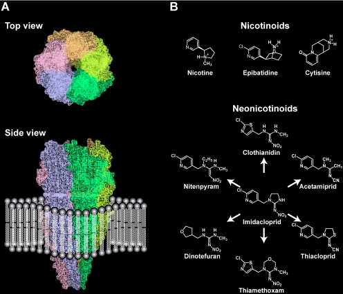
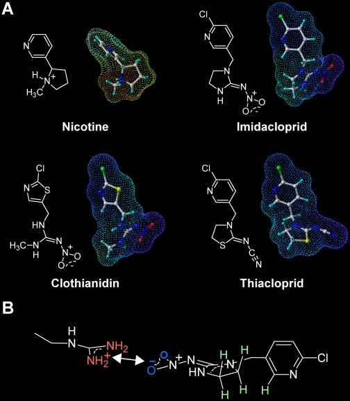
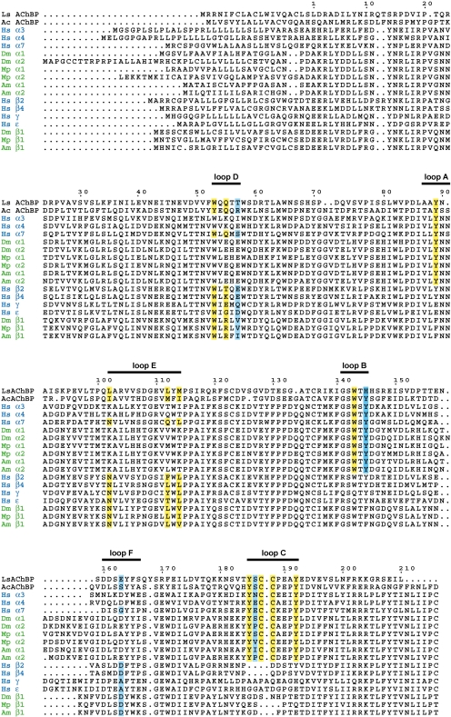
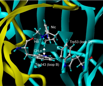
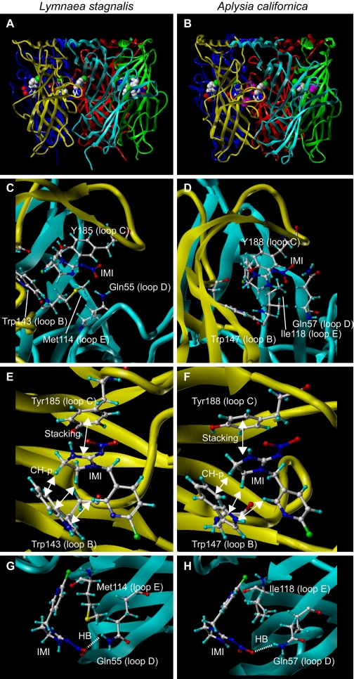
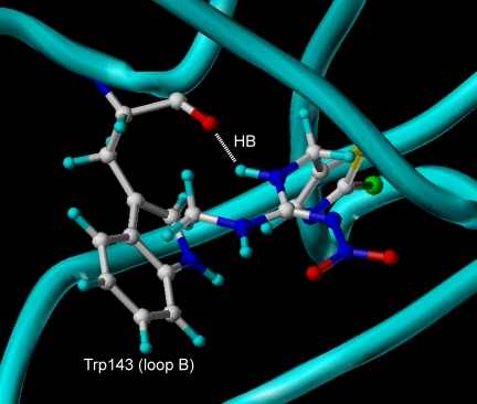
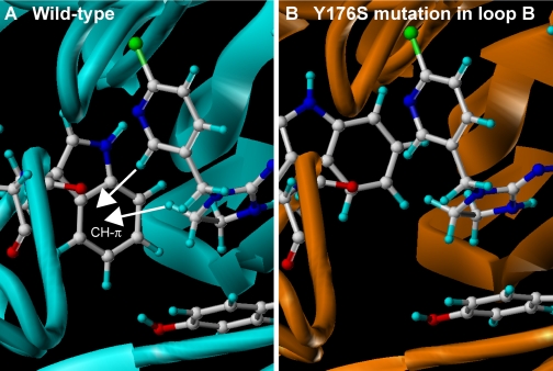
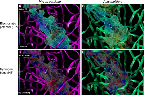
The nicotinic acetylcholine receptors (nAChRs) are targets for human and veterinary medicines as well as insecticides. Subtype-selectivity among the diverse nAChR family members is important for medicines targeting particular disorders, and pest-insect selectivity is essential for the development of safer, environmentally acceptable insecticides. Neonicotinoid insecticides selectively targeting insect nAChRs have important applications in crop protection and animal health. Members of this class exhibit strikingly diverse actions on their nAChR targets. Here we review the chemistry and diverse actions of neonicotinoids on insect and mammalian nAChRs. Electrophysiological studies on native nAChRs and on wild-type and mutagenized recombinant nAChRs have shown that basic residues particular to loop D of insect nAChRs are likely to interact electrostatically with the nitro group of neonicotinoids. In 2008, the crystal structures were published showing neonicotinoids docking into the acetylcholine binding site of molluscan acetylcholine binding proteins with homology to the ligand binding domain (LBD) of nAChRs. The crystal structures showed that 1) glutamine in loop D, corresponding to the basic residues of insect nAChRs, hydrogen bonds with the NO(2) group of imidacloprid and 2) neonicotinoid-unique stacking and CH-pi bonds at the LBD. A neonicotinoid-resistant strain obtained by laboratory-screening has been found to result from target site mutations, and possible reasons for this are also suggested by the crystal structures. The prospects of designing neonicotinoids that are safe not only for mammals but also for beneficial insects such as honey bees (Apis mellifera) are discussed in terms of interactions with non-alpha nAChR subunits.


Figures

Fig. 1.
Fig. 1.
The nAChR (A) and ligands (nicotinoids and neonicotinoids) (B). The model of the well characterized muscle nAChR was generated based on a PDB file of 2BG9 (Unwin, 2005) using Sybyl (version 7.1; Tripos Associates, Inc., St. Louis, MO).
Fig. 2.
Fig. 2.
Electrostatic potentials (EPs) surrounding nicotinoids and neonicotinoids (A), and interactions of imidacloprid with the arginine residue in insect nAChRs predicted by computational chemistry (B). In A, the EPs were calculated by the MNDO semiempirical molecular orbital method using the Sybyl (version 7.1; Tripos Associates, Inc.). Red dots indicate positive EPs, whereas blue dots indicate negative EPs. In B, the NO2 oxygens are highlighted in blue, whereas the protonated guanidine moiety of the arginine is highlighted in red. The hydrogens on the CH2-CH2 moiety in the imidazolidine ring and on the C2 carbon in the pyridine ring are colored light green because they are more electron-deficient than those on usual alkyl carbons. A part of these hydrogens form CH-π hydrogen bonds with the tryptophan ring in loop B in the crystal structures of acetylcholine binding proteins (see Fig. 4 for details).
Fig. 3.
Fig. 3.
Multiple sequence alignments of AChBPs with nicotinic acetylcholine receptor α and non-α subunits. Amino acids that have been shown to interact directly with nicotine and neonicotinoids are highlighted with a yellow background, whereas those indirectly determining neonicotinoid sensitivity are shown with a light blue background (The X residue in the YXCC motif is tentatively highlighted with blue). The six loops comprising the ligand binding domain are shown above the sequences. Ls, Lymnaea stagnalis; Ac, Aplysia californica; Hs, Homo sapiens; Dm, Drosophila melanogaster; Mp, Myzus persicae; Am, Apis mellifera.
Fig. 4.
Fig. 4.
Amino acids playing a critical role in the interactions with nicotine in the crystal structure of Ls acetylcholine binding protein (PDB, 1UV6). The picture was generated using Sybyl (version 7.1; Tripos Associates, Inc.). Note that nicotine is captured by a hydrogen bond between NH and the backbone of Trp143 as well as a CH-π interaction between N+CH2-H and the tryptophan ring. The backbone of the principal side donating loops A to C is colored yellow, whereas that of the complementary side donating loops D to F is colored cyan. Nic, nicotine; HB, hydrogen bond.
Fig. 5.
Fig. 5.
Amino acids interacting with a neonicotinoid imidacloprid in Ls- and Ac-AChBPs. A and B, side views of crystal structures of Ls- (A) and Ac- (B) AChBPs. All were prepared using Sybyl (version 7.1; Tripos Associates, Inc.). In A and B, imidacloprid and isopropyl alcohol (colored magenta) are generated in spheres to highlight. In C and D, amino acids interacting with imidacloprid in Ls- and Ac-AChBPs are shown, respectively. Irrespective of the mollusc species, a common mechanism is involved in the neonicotinoid recognition by the ligand binding domain of AChBPs. The main chain donating loops A to C is colored yellow, whereas the main chain giving loops D to F is shown cyan. In E (Ls-AChBP) and F (Ac-AChBP), amino acids from loops B and C are shown, whereas in G and H, those from loops D and E are shown in orientations facilitating view of interactions. Carbon, hydrogen, nitrogen, oxygen, and chlorine atoms are colored white, light blue, blue, red, and blue-green, respectively. IMI, imidacloprid; HB, hydrogen bond.
Fig. 6.
Fig. 6.
The ligand binding domain of Ls acetylcholine binding protein in complex with clothianidin. The figure was generated using Sybyl (version 7.1; Tripos Associates, Inc.). Carbon, hydrogen, nitrogen, oxygen, and chlorine and sulfur atoms of clothianidin are colored white, light blue, blue, red, blue-green, and yellow, respectively. Note that the NH of the guanidine moiety of clothianidin forms a hydrogen bond (HB) with the backbone C=O of Trp143 in loop B. Such a clothianidin-unique hydrogen bond may be involved in the super agonist actions of clothianidin and its analog on native D. melanogaster nAChRs (Brown et al., 2006a) as well as recombinant D. melanogaster Dα2/chicken β2 hybrid nAChRs expressed in X. laevis oocytes (Ihara et al., 2004).
Fig. 7.
Fig. 7.
LBD homology models of wild-type α2β1 nicotinic acetylcholine receptor from the peach potato aphid M. persicae (A) and Y176S mutant (B). Models were constructed according to Toshima et al. (2009). Modeling of the N-terminal region of M. persicae α2β1 and its Y176S mutant nAChRs was carried out using the molecular modeling software package Sybyl (version 7.3; Tripos Associates, Inc.) and the homology modeling software PDFAMS Ligands & Complex (version 2.1; In-Silico Sciences, Inc., Tokyo, Japan) in the ligand and complex mode. Both α2 and β1 subunits were aligned with the Ls-AChBP bound by imidacloprid (PDB code 2ZJU). In the second step, the three-dimensional structures of the wild-type and the Y176S mutant LBD-imidacloprid complexes were generated based on the sequence alignment and the coordinates of the AChBP and imidacloprid using the simulated annealing method (Kirkpatrick et al., 1983). The coordinates of imidacloprid were fixed during the simulated annealing. The receptor model constructed in this way was energy-minimized for 5000 iterations of conjugated gradients using the force field and partial charges of the molecular mechanics MMFF94 (Halgren 1999a,b) using Sybyl. Residues within a 10-Å radius of the centrally located imidacloprid, as well as imidacloprid itself, were treated as flexible entities except the C=N-NO2 moiety, which was fixed during energy minimization. In addition, residues within 10 to 15 Å radius of the ligand were considered rigid entities to speed up the computation. Other residues were ignored in energy minimization. Carbon, hydrogen, nitrogen, oxygen, and chlorine atoms of amino acids and imidacloprid are colored white, light blue, blue, red, and blue-green, respectively. Note that the CH-π hydrogen bonds with the tryptophan residue in loop B are reduced by this mutation, consistent with enhanced imidacloprid-resistance in pests.
Fig. 8.
Fig. 8.
Electrostatic (A and B) and hydrogen bonding (C and D) fields extended from the ligand binding site of the peach potato aphid M. persicae (A and C) and the honeybee A. mellifera (B and D). Models were constructed according to Toshima et al. (2009). In A and B, regions with high (positive) and low (negative) electrostatic potentials are colored red and blue, respectively. In C and D, the hydrogen-bond-accepting atoms such as nitrogens and oxygens are colored blue, whereas the hydrogen atoms attached to these hetero-atoms are colored red. In all, carbon, hydrogen, nitrogen, oxygen, and chlorine atoms of imidacloprid are colored white, light blue, blue, red, and blue-green, respectively. The grooves extending from the NO2 binding site in the pest and beneficial species nAChRs differ in terms of the size, electrostatic, and hydrogen bonding properties, which may lead to a generation of pest target-selective insecticides.

References

    1. Amiri S, Shimomura M, Vijayan R, Nishiwaki H, Akamatsu M, Matsuda K, Jones AK, Sansom MS, Biggin PC, and Sattelle DB (2008) A role for Leu118 of loop E in agonist binding to the α7 nicotinic acetylcholine receptor. Mol Pharmacol 73 1659-1667. - PubMed
    1. Arneric SP, Holladay M, and Williams M (2007) Neuronal nicotinic receptors: a perspective on two decades of drug discovery research. Biochem Pharmacol 74 1092-1101. - PubMed
    1. Bai D, Lummis S, Leicht W, Breer H, and Sattelle D (1991) Actions of imidacloprid and a related nitromethylene on cholinergic receptors of an identified insect motor neurone. Pestic Sci 33 197-204.
    1. Bertrand D, Ballivet M, Gomez M, Bertrand S, Phannavong B, and Gundelfinger ED (1994) Physiological properties of neuronal nicotinic receptors reconstituted from the vertebrate β2 subunit and Drosophila a subunits. Eur J Neurosci 6 869-875. - PubMed
    1. Bocquet N, Nury H, Baaden M, Le Poupon C, Changeux JP, Delarue M, and Corringer PJ (2009) X-ray structure of a pentameric ligand-gated ion channel in an apparently open conformation. Nature 457 111-114. - PubMed

Publication types