. 2009 Mar 25;29(12):3843-51.
doi: 10.1523/JNEUROSCI.5842-08.2009.

Cisplatin-induced hair cell death requires STAT1 and is attenuated by epigallocatechin gallate

Affiliations

Cisplatin-induced hair cell death requires STAT1 and is attenuated by epigallocatechin gallate

Nicole C Schmitt et al. J Neurosci. .

Abstract

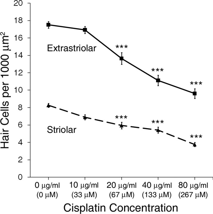
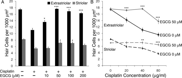
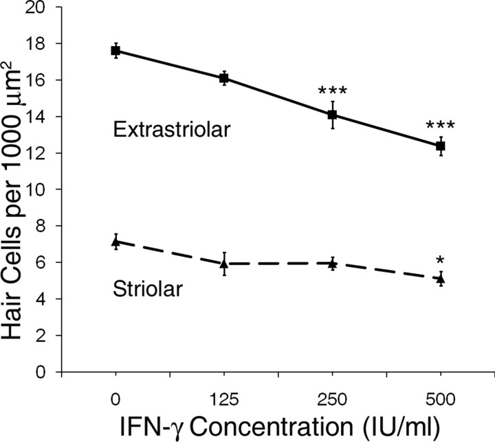
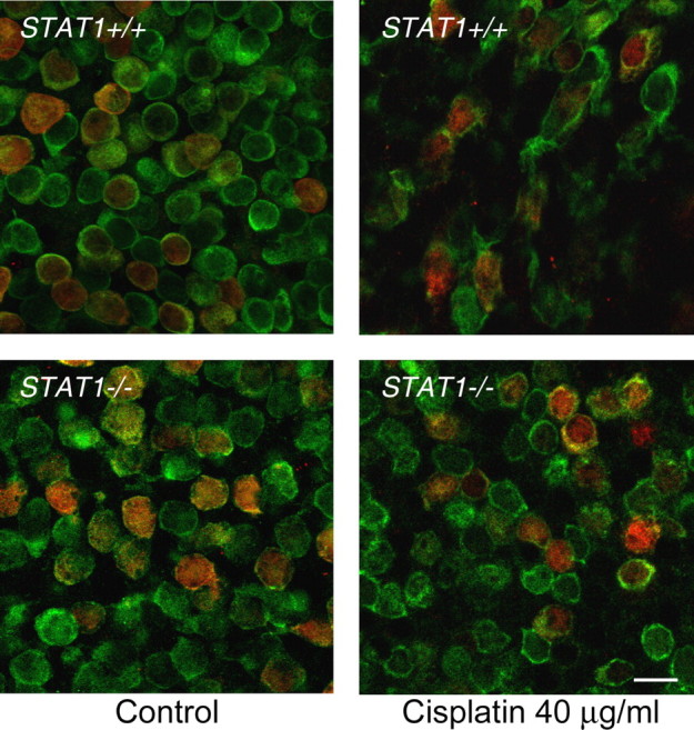
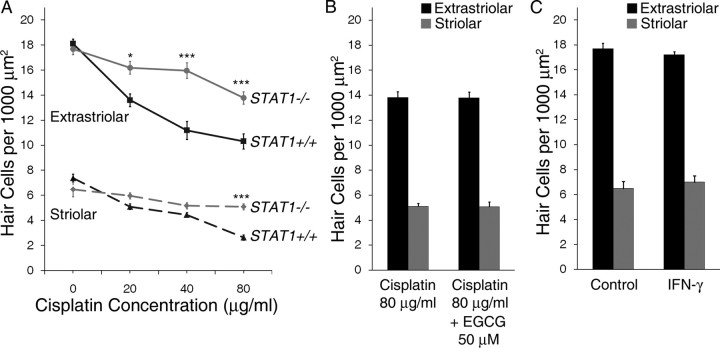
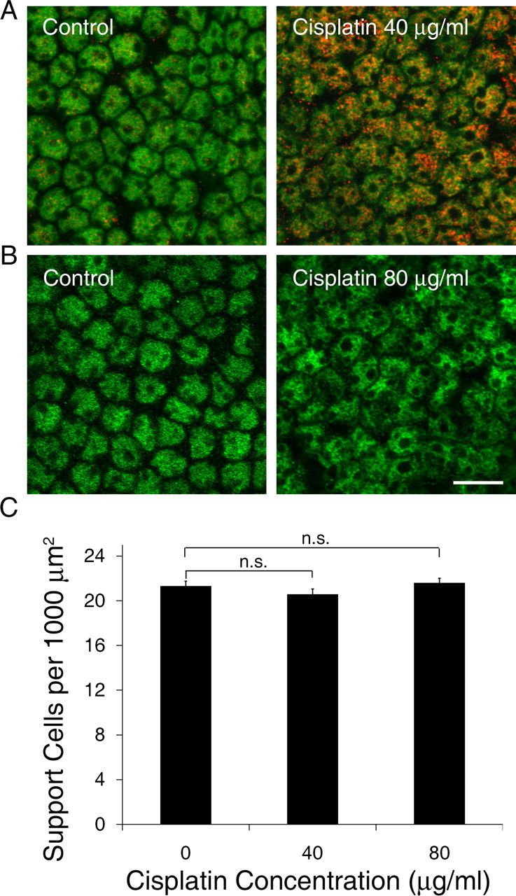
Cisplatin is a chemotherapy drug that frequently causes auditory impairment due to the death of mechanosensory hair cells. Cisplatin ototoxicity may result from oxidative stress, DNA damage, and inflammatory cytokines. The transcription factor STAT1, an important mediator of cell death, can regulate all of these processes in other cell types. We used cultured utricles from mature Swiss Webster mice to investigate the role of STAT1 in cisplatin-induced hair cell death. We show that STAT1 phosphorylation is an early event in both hair cells and support cells after exposure of utricles to cisplatin. STAT1 phosphorylation peaked after 4 h of cisplatin exposure and returned to control levels by 8 h of exposure. The STAT1 inhibitor epigallocatechin gallate (EGCG) attenuated STAT1 phosphorylation in cisplatin-treated utricles and resulted in concentration-dependent increases in hair cell survival at 24 h postexposure. Furthermore, we show that utricular hair cells from STAT1-deficient mice are resistant to cisplatin toxicity. EGCG failed to provide additional protection from cisplatin in STAT1-deficient mice, further supporting the hypothesis that the protective effects of EGCG are due to its inhibition of STAT1. Treatment with IFN-gamma, which also causes STAT1 activation, also induced hair cell death in wild-type but not STAT1-deficient mice. These results show that STAT1 is required for maximal cisplatin-induced hair cell death in the mouse utricle and suggest that treatment with EGCG may be a useful strategy for prevention of cisplatin ototoxicity.


Figures

Figure 1.
Figure 1.
Cisplatin treatment resulted in concentration-dependent decreases in hair cell survival (mean ± 1 SEM, p < 0.001). The concentration–response relationships of extrastriolar hair cells (solid line, squares) and striolar hair cells (dashed line, triangles) were determined in utricles treated for 24 h with increasing concentrations of cisplatin. Equivalent concentrations of cisplatin in μm are given in parentheses for comparison. Hair cell density was determined as the average number of hair cells per 1000 μm2 area. ***p < 0.001 compared with control.
Figure 2.
Figure 2.
Serine phosphorylation of STAT1 is an early event after IFN-γ and cisplatin exposure. Mouse utricles were cultured for 1, 4, or 8 h in control medium (A, E, I), with IFN-γ as a positive control (B, F, J), with cisplatin at 40 μg/ml (133 μm; C, G, K), or with cisplatin at 40 μg/ml plus the STAT1 inhibitor EGCG at 50 μm (D, H, L). Hair cells were labeled with an antibody against calmodulin (green) and an antibody against pSTAT1Ser (red). Nuclei were counterstained with Hoechst 33258 (blue). Each inset shows an enlargement of one hair cell with its nucleus outlined (white dotted line). Very little pSTAT1Ser was noted in hair cells from control utricles (A, E, I). A faint pSTAT1Ser label was noted in the IFN-γ and cisplatin-treated utricles at 1 h (B, C), which peaked at 4 h (F, G) and was diminished by 8 h (J, K). In utricles treated with cisplatin plus EGCG, the pSTAT1Ser label consistently appeared diminished compared with utricles treated with cisplatin alone at 1 and 4 h (D, H). Examples shown are most representative of 6–8 utricles in each group from three to five independent experiments. Scale bars represent 10 μm.
Figure 3.
Figure 3.
EGCG reduces hair cell loss and morphologic damage after cisplatin treatment. Utricles were cultured in control medium or a moderate concentration of cisplatin for 24 h with or without the STAT1 inhibitor EGCG. Confocal photomicrographs were taken at the junction of extrastriolar hair cells (green, labeled for calmodulin) and striolar hair cells (red and green, labeled for calbindin and calmodulin, respectively). Treatment with cisplatin alone resulted in hair cell loss and morphologic damage, including cellular shrinkage or swelling. When treated with cisplatin plus EGCG, utricular hair cells demonstrated near-normal density and morphology. Examples shown are representative utricles from at least three independent experiments. Scale bar represents 10 μm.
Figure 4.
Figure 4.
EGGC protects utricular hair cells from cisplatin. A, Utricles were treated for 24 h with or without a moderate concentration of cisplatin (40 μg/ml) and increasing concentrations of EGCG, which resulted in concentration-dependent increases in hair cell survival up to the optimal concentration of 50 μm EGCG (mean + 1 SEM, p < 0.001 for extrastriolar, p < 0.01 for striolar, n = 7–10 utricles per group). B, Utricles were treated for 24 h with increasing concentrations of cisplatin with or without the optimal concentration of EGCG (50 μm, based on results in A). Density of both extrastriolar hair cells (solid lines) and striolar hair cells (dashed lines) was increased by EGCG (mean ± 1 SEM, p < 0.001, n = 7–10 utricles per group). *p < 0.05, **p < 0.01, and ***p < 0.001 versus same dose of cisplatin without EGCG. Equivalent μm concentrations of cisplatin are shown in Figure 1.
Figure 5.
Figure 5.
Effects of IFN-γ on hair cell survival. Utricles from wild-type Swiss Webster mice were treated with increasing concentrations of IFN-γ (0 – 500 IU/ml) for 24 h, which resulted in a concentration-dependent decrease in survival of both extrastriolar hair cells (solid line) and striolar hair cells (dashed line) (mean ± 1 SEM, p < 0.001 and p < 0.05 for extrastriolar and striolar, respectively; n = 7–9 utricles per group). *p < 0.05, ***p < 0.001 compared with control.
Figure 6.
Figure 6.
Utricles from STAT1−/− mice are resistant to hair cell loss and morphologic damage after cisplatin treatment. Utricles from STAT1+/+ and STAT1−/− mice were cultured in control medium or with a moderate concentration of cisplatin for 24 h. Confocal photomicrographs were taken at the junction of extrastriolar hair cells (green, labeled for calmodulin) and striolar hair cells (red and green, labeled for calbindin and calmodulin, respectively). Treatment of utricles from STAT1+/+ mice with cisplatin resulted in decreased hair cell density and substantial morphologic damage, including hair cell swelling or shrinkage. Utricles from STAT1−/− mice treated with cisplatin demonstrated near-normal density and morphology. Examples shown are representative utricles from three independent experiments. Scale bar represents 10 μm.
Figure 7.
Figure 7.
Utricles from STAT1−/− mice are protected from cisplatin- and IFN-γ-induced hair cell death and are unaffected by EGCG. A, Utricles from STAT1+/+ and STAT1−/− mice were treated with increasing concentrations of cisplatin for 24 h. Death of both extrastriolar hair cells (solid lines) and striolar hair cells (dashed lines) was significantly decreased in utricles from STAT1−/− mice compared with utricles from STAT1+/+ mice treated with the same concentrations of cisplatin (mean ± 1 SEM, p < 0.001, n = 5–6 utricles per group). *p < 0.05, ***p < 0.001 vs same dose of cisplatin in utricles from STAT1+/+ mice. Equivalent μm concentrations of cisplatin are shown in Figure 1. B, Utricles from STAT1−/− mice were treated with a high concentration of cisplatin alone or cisplatin plus EGCG for 24 h. The addition of EGCG failed to increase hair cell survival (mean + 1 SEM, p > 0.9, n = 5–6 utricles per group). C, Utricles from STAT1−/− mice were cultured in control medium or a high concentration of IFN-γ (500 IU/ml) for 24 h. IFN-γ did not affect hair cell survival versus utricles treated with control medium (mean + 1 SEM, p > 0.4, n = 5–6 utricles per group).
Figure 8.
Figure 8.
Effect of cisplatin on support cells. A, Utricles were treated with control medium or a moderate concentration of cisplatin for 4 h and labeled with pSTAT1Ser (red) and SOX2 (green) to visualize support cell nuclei. Support cells from control utricles demonstrated scant, punctate pSTAT1Ser (left), whereas pSTAT1Ser was abundant in nuclei of support cells from cisplatin-treated utricles (right). B, Utricles were treated with control medium or a high concentration of cisplatin for 24 h and labeled with SOX2 to visualize support cells. Cisplatin treatment resulted in mild disorganization but similar overall density of support cells (right). Scale bar corresponds to all panels and represents 10 μm. C, Utricles were treated with control medium or cisplatin for 24 h and support cell density was quantified. Cisplatin treatment did not result in a loss of support cells within 24 h (mean + 1 SEM, p > 0.2, n = 7–8 utricles per group). Equivalent μm concentrations of cisplatin are shown in Figure 1.

References

    1. Barton C, Davies D, Balkwill F, Burke F. Involvement of both intrinsic and extrinsic pathways in IFN-γ-induced apoptosis that are enhanced with cisplatin. Eur J Cancer. 2005;41:1474–1486. - PubMed
    1. Battle TE, Frank DA. The role of STATs in apoptosis. Curr Mol Med. 2002;2:381–392. - PubMed
    1. Bodmer D, Brors D, Bodmer M, Pak K, Ryan AF. Fas ligand expression in the organ of Corti. Audiol Neurootol. 2003;8:243–249. - PubMed
    1. Chen D, Milacic V, Chen MS, Wan SB, Lam WH, Huo C, Landis-Piwowar KR, Cui QC, Wali A, Chan TH, Dou QP. Tea polyphenols, their biological effects and potential molecular targets. Histol Histopathol. 2008;23:487–496. - PMC - PubMed
    1. Chiu LL, Cunningham LL, Raible DW, Rubel EW, Ou HC. Using the zebrafish lateral line to screen for ototoxicity. J Assoc Res Otolaryngol. 2008;9:178–190. - PMC - PubMed

Publication types

MeSH terms