. 2009 May 7;459(7243):103-7.
doi: 10.1038/nature07859. Epub 2009 Mar 25.

Haematopoietic stem cells depend on Galpha(s)-mediated signalling to engraft bone marrow

Affiliations

Haematopoietic stem cells depend on Galpha(s)-mediated signalling to engraft bone marrow

Gregor B Adams et al. Nature. .

Abstract

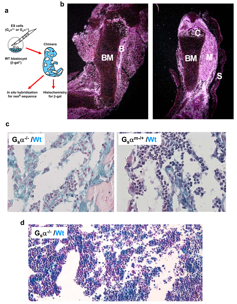
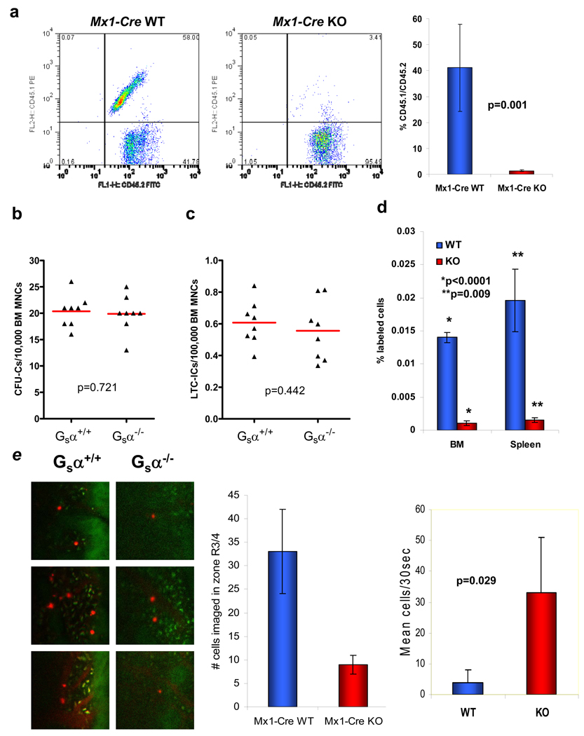
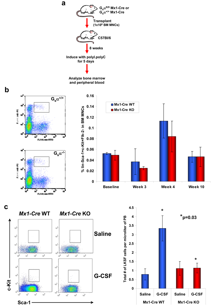
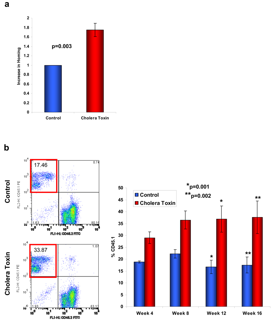
Haematopoietic stem and progenitor cells (HSPCs) change location during development and circulate in mammals throughout life, moving into and out of the bloodstream to engage bone marrow niches in sequential steps of homing, engraftment and retention. Here we show that HSPC engraftment of bone marrow in fetal development is dependent on the guanine-nucleotide-binding protein stimulatory alpha subunit (Galpha(s)). HSPCs from adult mice deficient in Galpha(s) (Galpha(s)(-/-)) differentiate and undergo chemotaxis, but also do not home to or engraft in the bone marrow in adult mice and demonstrate a marked inability to engage the marrow microvasculature. If deleted after engraftment, Galpha(s) deficiency did not lead to lack of retention in the marrow, rather cytokine-induced mobilization into the blood was impaired. Testing whether activation of Galpha(s) affects HSPCs, pharmacological activators enhanced homing and engraftment in vivo. Galpha(s) governs specific aspects of HSPC localization under physiological conditions in vivo and may be pharmacologically targeted to improve transplantation efficiency.


Figures

Figure 1
Figure 1. Gsα is required for HSPC engraftment of bone marrow in development
(a) Chimeric mice were created by injection of Gsα−/− or Gsα−/+ embryonic stem cells into wild-type or β-galactosidase transgenic blastocysts. At E17.5, the mice were killed and the organs were probed for Gsα−/− cells (using a neo probe). (b) In situ hybridization of femur and tibia is shown. Bone marrow (BM), bone (B), muscle (M), skin (S) and chondrocytes (C) are indicated. (c) Mice were assessed by histochemistry for β-galactosidase (blue stain) in Gsα−/− (left panel) or Gsαm−/+ (right panel) chimeric animal bone marrow. (d) Histochemical analysis of β-galactosidase expression in fetal liver from E13.5 Gsα−/− chimeric mice.
Figure 2
Figure 2. Gsα is required for HSPC engraftment of bone marrow in adults
Gsα was conditionally deleted in hematopoietic cells of 6-week old Mx1-Cre Gsαfl/fl mice and cells were then assessed for engraftment capability. (a) Representative flow cytometric analysis at 16 weeks and collated data of engraftment of Mx1-Cre WT (Gsα+/+) or Mx1-Cre KO (Gsα−/−) cells in competitive repopulation assay (n=8 from 2 independent experiments; error bars=s.e.m.). BM MNCs were assessed for their in vitro growth potential using (b) CFU-C and (c) LTC-IC assays. (d) In vivo homing to WT bone marrow and spleen of Mx1-Cre WT and KO LKS+ cells (n=3; error bars represent s.e.m.). (e) Intravital imaging of calvarium bone marrow of WT recipient mice injected by tail vein with Mx1-Cre WT or KO LKS+ cells stained with DiD. Representative images (red: DiD, green: autofluorescence) and quantitation (graphs) of the number of cells visualized over a 30 second interval at ~35 min. post-infusion to be stably localized or circulating is shown (n=3; error bars=s.d.).
Figure 3
Figure 3. Gsα signalling is not required for retention of the HSPCs in the BM, but does influence mobilization by G-CSF
(a) Diagrammatic representation of the procedure to assess Gsα signalling in HSPC BM retention. Mx1-Cre Gsα+/+ or Gsαfl/fl cells were injected into wild-type mice; after 8 weeks, deletion of the Gsα gene was induced by polyI.polyC and BM and PB evaluated. (b) Retention of primitive LKS Flk-2 Mx1-Cre WT or KO cells in the bone marrow. Representative flow cytometric analysis at week 10 and mean collated data shown (n=5; error bars=s.e.m.). (c) Representative flow cytometric plots (left) and quantitation (right) of HSPC mobilization into the peripheral circulation following 5 days of G-CSF. Sca-1 and c-Kit expression of gated LinFlk-2 cells is shown (n=5; error bars=s.e.m.).
Figure 4
Figure 4. Pharmacological modulation of Gsα affects homing and engraftment of primitive wild-type BM MNCs
(a) Primitive lin BM MNCs pre-treated with cholera toxin or mock treated for 1 hour ex vivo were assessed for their in vivo homing potential (n=3 from 3 individual experiments; error bars represent s.e.m.). (b) Engraftment potential in a competitive transplant model (n=9 from 2 individual experiments; error bars represent s.e.m.).

References

    1. Mikkola HKA, Orkin SH. The journey of developing hematopoietic stem cells. Development. 2006;133:3733–3744. - PubMed
    1. Wright DE, Wagers AJ, Gulati AP, Johnson FL, Weissman IL. Physiological migration of hematopoietic stem and progenitor cells. Science. 2001;294:1933–1936. - PubMed
    1. Mazo IB, et al. Hematopoietic progenitor cell rolling in bone marrow microvessels: parallel contributions by endothelial selectins and vascular cell adhesion molecule 1. J. Exp. Med. 1998;188:465–474. - PMC - PubMed
    1. Papayannopoulou T, Priestley GV, Nakamoto B, Zafiropoulos V, Scott LM. Molecular pathways in bone marrow homing: dominant role of β4β1 over β2-integrins and selectins. Blood. 2001;98:2403–2411. - PubMed
    1. Lapidot T, Dar A, Kollet O. How do stem cells find their way home? Blood. 2005;106:1901–1910. - PubMed

Methods References

    1. Calvi LM, et al. Osteoblastic cells regulate the haematopoietic stem cell niche. Nature. 2003;425:841–846. - PubMed
    1. Cheng T, et al. Hematopoietic stem cell quiescence maintained by p21cip1/waf1. Science. 2000;287:1804–1808. - PubMed
    1. Stier S, Cheng T, Dombkowski D, Carlesso N, Scadden DT. Notch1 activation increases hematopoietic stem cell self-renewal in vivo and favors lymphoid over myeloid lineage outcome. Blood. 2002;99:2369–2378. - PubMed

Publication types

MeSH terms

Substances