. 2009 Apr 22;28(8):1131-41.
doi: 10.1038/emboj.2009.75. Epub 2009 Mar 26.

Differential regulation of homologous recombination at DNA breaks and replication forks by the Mrc1 branch of the S-phase checkpoint

Affiliations

Differential regulation of homologous recombination at DNA breaks and replication forks by the Mrc1 branch of the S-phase checkpoint

Constance Alabert et al. EMBO J. .

Abstract

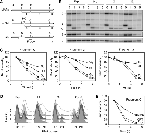
The Rad52 pathway has a central function in the recombinational repair of chromosome breaks and in the recovery from replication stress. Tolerance to replication stress also depends on the Mec1 kinase, which activates the DNA replication checkpoint in an Mrc1-dependent manner in response to fork arrest. Although the Mec1 and Rad52 pathways are initiated by the same single-strand DNA (ssDNA) intermediate, their interplay at stalled forks remains largely unexplored. Here, we show that the replication checkpoint suppresses the formation of Rad52 foci in an Mrc1-dependent manner and prevents homologous recombination (HR) at chromosome breaks induced by the HO endonuclease. This repression operates at least in part by impeding resection of DNA ends, which is essential to generate 3' ssDNA tails, the primary substrate of HR. Interestingly, we also observed that the Mec1 pathway does not prevent recombination at stalled forks, presumably because they already contain ssDNA. Taken together, these data indicate that the DNA replication checkpoint suppresses genomic instability in S phase by blocking recombination at chromosome breaks and permitting helpful recombination at stalled forks.


Figures

Figure 1
Figure 1
Genotoxic agents differentially affect the formation of Rad52 foci in S phase. (A) Overview of the assay. Wild-type cells (PP534) expressing Rad52–GFP were arrested in G1 with α-factor for 120 min. Half of the culture was exposed to the radiomimetic agent Zeocin (ZEO) for 30 min to generate DSBs. Cells treated with or without Zeocin were either maintained in G1 or released into S phase for the indicated time. (B) The percentage of G1- and S-phase cells forming spontaneous and Zeocin-induced Rad52 foci was scored as described in Materials and Methods. Representative images of cells showing Rad52–GFP foci are shown. (C) Percentage of wild-type cells forming Rad52 foci in response to genotoxic drugs. Rad52–GFP cells were arrested in G1 and were released into S phase in the absence of drugs (untreated) or in the presence of 200 mM hydroxyurea (HU), 0.033% methyl methanesulphonate (MMS), 20 μM camptothecin (CPT) or 100 μg/ml Zeocin (ZEO). The fraction of cells showing Rad52–GFP foci was determined for the indicated time after release from α-factor. (D, E) Cells arrested at the G1/S transition are competent to form Rad52 foci. Thermosensitive cdc7-4 cells (PP544) expressing Rad52–GFP were arrested in G1 with α-factor for 120 min and exposed to 100 μg/ml Zeocin for 30 min in the presence of α-factor. Cells were then released into S phase at the permissive (24°C) or restrictive (37°C) temperature for the cdc7-4 mutation and the percentage of cells forming Rad52 foci was scored 90 min after release from G1. Flow cytometry analysis indicates that although cdc7-4 cells are blocked at the G1/S transition (D), they are able to assemble Rad52 foci (E).
Figure 2
Figure 2
HU and MMS prevent the assembly of Rad52 foci at DSBs. (A) Wild-type cells (PP534) expressing Rad52–GFP were arrested in G1, exposed to Zeocin as described in Figure 1 and released into S phase for 90 min in fresh medium containing no drug (S), 200 mM HU, 0.033% MMS or 20 μM CPT. The fraction of cells showing Rad52 foci was scored. (B) Wild-type cells expressing Rad52–GFP were arrested in G1 for 120 min and cells were maintained in G1 for another 30 min in the presence or absence of 100 μg/ml Zeocin (grey box). Cells were then released into S phase and HU was added to the culture at the indicated time (white boxes). (C) Percentage of cells presenting Rad52–GFP foci. (D) PFGE analysis of chromosome mobility in Zeocin-treated cells exposed to HU at the indicated time points after release from G1. Averaged values for a representative set of chromosomes are shown. (E) cdc7-4 mutants (PP544) were arrested in G1 for 120 min at 24°C and exposed to 100 μg/ml Zeocin for another 60 min at 24°C. Cells were then released into S phase for 90 min in the presence (ZEO+HU) or the absence (ZEO) of 200 mM hydroxyurea. The percentage of cells presenting Rad52 foci is shown.
Figure 3
Figure 3
Cells exposed to HU or MMS are unable to repair HO-induced DSBs. (A) Overview of the assay. Early log-phase cultures of wild-type cells (PP723) bearing a GAL-HO construct were exposed to 200 mM HU for 4 h, 0.033% MMS for 2 h or 20 μM CPT for 3 h prior to HO induction (see Supplementary Figure 1B for DNA content profiles). Expression of the HO endonuclease was induced for 60 min with 2% galactose and repressed with 2% glucose. Genomic DNA was extracted at the indicated time points and was digested with StyI (S). The smallest of the two StyI fragments (*) generates fragment C (cut) upon cleavage with HO. Homologous recombination with HMLα generates fragment R (repair). (B) Southern blot analysis of DSB repair in untreated cells (ctrl) or in cells exposed to HU, MMS or CPT. (C) Relative intensity of the repair band in untreated control cells and in cells exposed to HU, MMS and CPT.
Figure 4
Figure 4
HU and MMS impede resection of HO-induced DSB. (AC) Exponentially growing (Exp.) wild-type cells (PP1123) bearing a GAL-HO construct were arrested in G1 with α-factor, in G2 with 15 μg/ml nocodazole or in S phase with 200 mM HU. HO expression was induced as described above and the resection of HO-induced DSB at the MATa locus was monitored by Southern blot hybridization. The rate of disappearance of the HO-cut band (fragment C) and of adjacent StyI fragments (2 and 3) was expressed for each time point as a percentage of the intensity of the initial band. The intensity of resection bands was normalized to the intensity of the RAD9 gene (*). (D) Flow cytometry analysis of DNA content in exponentially growing cells and in cells arrested in G1, G2 or with HU. (E) Southern blot analysis of the resection rate of HO-induced DSB in the presence of 0.033% MMS or 20 μM CPT.
Figure 5
Figure 5
Mrc1 suppresses the formation HR foci and ssDNA overhangs in response to replication stress. (A) Wild-type (PP534), mrc1Δ (PP388), rad9Δ (PP1154), mrc1AQ (PP471) and mrc1AQ rad9Δ (PP1064) cells were arrested in G1 and were exposed or not to Zeocin (ZEO) as indicated in Figure 2A. Cells were then released into S phase for 90 min in the presence or absence of 200 mM HU. The percentage of cells containing Rad52–GFP foci is indicated. (B) Percentage of wild-type (PP534) and mrc1Δ (PP388) cells forming Zeocin-induced Rad52 foci in S phase after exposure to HU, MMS or CPT. Cells were arrested in G1, treated with Zeocin and were released into S phase for 90 min in the presence (HU, MMS and CPT) or absence (S) of drugs as described in Figure 1B. (C) Analysis of DSB resection at MATα in untreated mrc1Δ (PP736) cells (Exp.) or in cells exposed to HU, MMS or CPT. Experiment was performed as described in Figure 4. (D, E) Resection rate at the HO-cut band (fragment C) and at the adjacent StyI fragment (fragment 2) in untreated mrc1Δ cells (Exp.) and in cells exposed to drugs (HU, MMS and CPT). The ratio of intensity of fragments in the presence or absence of HU is indicated for wild-type and mrc1Δ cells.
Figure 6
Figure 6
Rad51 is required for the recovery of MMS-induced replication fork stalling. Wild-type (PP633) and rad51Δ (PP484) cells were arrested in G1 with α-factor and released synchronously into S phase in the presence of 400 μg/ml BrdU and 0.033% MMS for 60 min. Cells were then collected and resuspended in fresh medium containing BrdU for the indicated time after MMS release (t=0). (A) PFGE analysis of chromosome mobility in wild-type and rad51Δ cells after release from MMS arrest. (B) Quantitation of chromosome mobility during release from MMS. (C) DNA fibres from wild-type and rad51Δ cells treated as indicated above were analysed by DNA combing. Representative DNA fibres from MMS-treated cells are shown. Red: DNA; green: BrdU; white: BrdU channel alone; brackets: unreplicated gaps. Scale bar is 50 kb. (D) Quantitation of the percentage of replication and the number of unreplicated gaps per megabyte of genomic DNA in wild-type and rad51Δ cells released from MMS arrest. (E) 2D gel analysis of replication intermediates in wild-type, rad51Δ and sgs1Δ cells exposed for 60 min to 0.1% MMS. 2D gel electrophoresis was performed as described, using a probe against the early origin ARS305 (Tourriere et al, 2005). X, X-spike; B, bubble arc; Y, Y arc. Closed and open arrowheads point to the presence or the absence of X-spikes, respectively.
Figure 7
Figure 7
Differential regulation of homologous recombination at DNA double-strand breaks and stalled forks under replication stress. (A) DSBs are efficiently repaired by homologous recombination (HR) during S phase after the formation of 3′ ssDNA overhangs. (B) In the presence of HU or MMS, stalled replication forks induce the activation of the DNA replication checkpoint in an Mrc1-dependent manner through the recruitment of the Mec1–Ddc2 complex on RPA-coated ssDNA. Hyperphosphorylated Rad53 functions in trans to prevent HR at DSBs, presumably by blocking the formation of ssDNA tails. In contrast, sister-chromatid recombination occurs at stalled forks, which already contain ssDNA (see text for details). SCC, sister-chromatid cohesion.

References

    1. Alcasabas AA, Osborn AJ, Bachant J, Hu F, Werler PJ, Bousset K, Furuya K, Diffley JF, Carr AM, Elledge SJ (2001) Mrc1 transduces signals of DNA replication stress to activate Rad53. Nat Cell Biol 3: 958–965 - PubMed
    1. Alvino GM, Collingwood D, Murphy JM, Delrow J, Brewer BJ, Raghuraman MK (2007) Replication in hydroxyurea: it's a matter of time. Mol Cell Biol 27: 6396–6406 - PMC - PubMed
    1. Aylon Y, Liefshitz B, Kupiec M (2004) The CDK regulates repair of double-strand breaks by homologous recombination during the cell cycle. EMBO J 23: 4868–4875 - PMC - PubMed
    1. Bashkirov VI, King JS, Bashkirova EV, Schmuckli-Maurer J, Heyer WD (2000) DNA repair protein Rad55 is a terminal substrate of the DNA damage checkpoints. Mol Cell Biol 20: 4393–4404 - PMC - PubMed
    1. Boddy MN, Shanahan P, McDonald WH, Lopez-Girona A, Noguchi E, Yates IJ, Russell P (2003) Replication checkpoint kinase Cds1 regulates recombinational repair protein Rad60. Mol Cell Biol 23: 5939–5946 - PMC - PubMed

Publication types

MeSH terms