

Myocardial 3D strain calculation by combining cine displacement encoding with stimulated echoes (DENSE) and cine strain encoding (SENC) imaging
- PMID: 19322795
- PMCID: PMC2736095
- DOI: 10.1002/mrm.21984
Myocardial 3D strain calculation by combining cine displacement encoding with stimulated echoes (DENSE) and cine strain encoding (SENC) imaging
Abstract
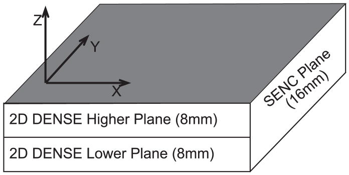
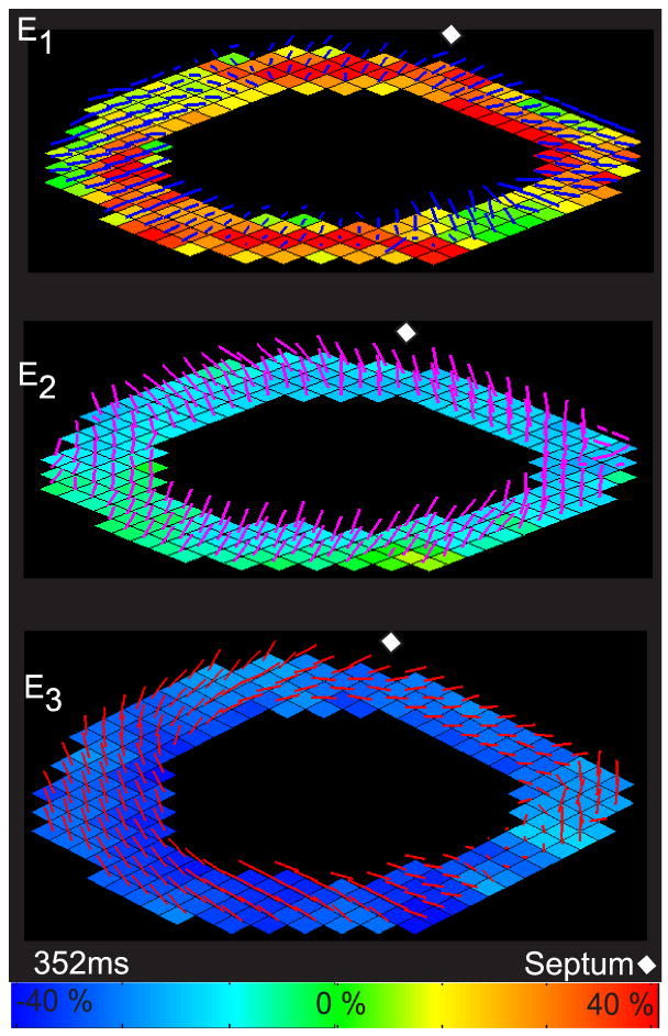
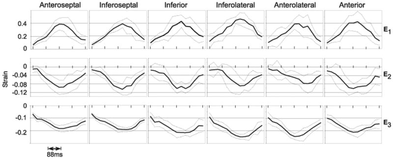
Three-dimensional (3D) strain maps of the myocardium provide a coordinate-system-independent quantification of myocardial deformation and kinematics. We combine two MRI techniques, displacement encoding with stimulated echoes (DENSE) and strain encoding (SENC), to fully formulate a 3D strain map in a single slice of myocardium. The method utilizes 2D DENSE in-plane displacement measurements in two adjacent slices in conjunction with a single SENC through-plane strain measure to calculate the 3D strain tensor. Six volunteers were imaged and the technique demonstrated 3D strain measures in all volunteers that are consistent with those reported in the literature from 3D tagging. The mean peak strain (+/- standard deviation [SD]) for six healthy volunteers for the first, second, and third principal strains are 0.42 +/-0.11, -0.10 +/-0.03, and -0.21 +/-0.02, respectively. These results show that this technique is capable of reliably quantifying 3D cardiac strain.
(c) 2009 Wiley-Liss, Inc.
Figures
References
-
- Kim D, Gilson WD, Kramer CM, Epstein FH. Myocardial Tissue Tracking with Two-dimensional Cine Displacement-encoded MR Imaging: development and Initial Evaluation. Radiology. 2004;230:862–871. - PubMed
-
- Osman NF, Sampath S, Atalar E, Prince JL. Imaging Longitudinal Cardiac Strain on Short-Axis Images Using Strain-Encoded MRI. Magnetic Resonance in Medicine. 2001;46:324–334. - PubMed
-
- Zerhouni EA, Parish DM, Rogers WJ, Yang A, Shapiro EP. Human Heart: Tagging with MR Imaging – A new Method for Noninvasive Assement of Myocardial Motion. Cardiac Radiology. 1988;169:59–63. - PubMed
-
- Axel L, Dougherty L. MR Imaging of Motion with Spatial Modulation of Magnetization. Radiology. 1989;171:841–845. - PubMed
Publication types
MeSH terms
Grants and funding
LinkOut - more resources
Full Text Sources
Other Literature Sources
