

Activated leukocyte cell adhesion molecule: a novel biomarker for breast cancer
- PMID: 19322904
- PMCID: PMC3743675
- DOI: 10.1002/ijc.24292
Activated leukocyte cell adhesion molecule: a novel biomarker for breast cancer
Abstract
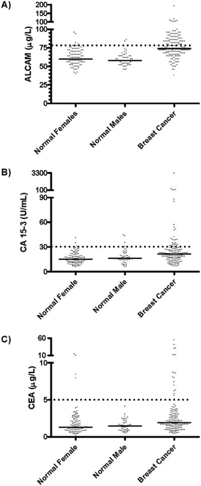
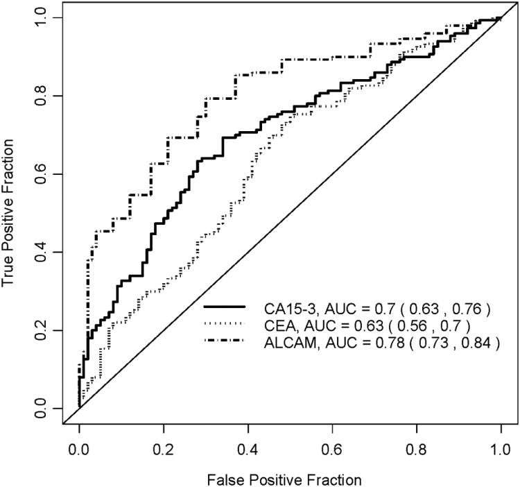
Activated leukocyte cell adhesion molecule (ALCAM) has been implicated in tumorigenesis. Our goal was to examine the levels of ALCAM, in addition to the classical breast cancer tumor markers carbohydrate antigen 15-3 (CA15-3) and carcinoembryonic antigen (CEA), in serum by quantitative enzyme-linked immunosorbent assay for diagnosis in breast cancer patients. The 3 proteins were measured in serum of 100 healthy women, 50 healthy men and 150 breast carcinoma patients. The diagnostic sensitivity and specificity of the tests were calculated and the association of serum marker concentrations with various clinicopathologic variables was examined using nonparametric Kruskal-Wallis tests. Receiver operating characteristic (ROC) curves were used to evaluate the diagnostic performance of the biomarkers. ALCAM, with area under the curve (AUC) of 0.78 [95% CI: 0.73, 0.84] outperformed CA15-3 (AUC = 0.70 [95% CI: 0.64, 0.76]) and CEA (AUC= 0.63 [95% CI: 0.56, 0.70]). The incremental values of AUC for ALCAM over that for CA15-3 were statistically significant (Delong test, p < 0.05). Combining CA15-3 and ALCAM yielded a ROC curve with an AUC of 0.81 (95% CI [0.75, 0.87]). Serum ALCAM appears to be a new biomarker for breast cancer and may have value for disease diagnosis.
Figures
Similar articles
-
Thioredoxin 1 as a serum marker for breast cancer and its use in combination with CEA or CA15-3 for improving the sensitivity of breast cancer diagnoses.BMC Res Notes. 2014 Jan 6;7:7. doi: 10.1186/1756-0500-7-7. BMC Res Notes. 2014. PMID: 24393391 Free PMC article. Clinical Trial.
-
Assessment of serum microRNA-21 and miRNA-205 as diagnostic markers for stage I and II breast cancer in Indian population.Indian J Cancer. 2024 Apr 1;61(2):290-298. doi: 10.4103/ijc.IJC_187_20. Epub 2023 Jul 17. Indian J Cancer. 2024. PMID: 38090957
-
The Clinical Significance of sICAM-1 in Differentiating Benign Breast Lesions from Breast Cancer.Ann Clin Lab Sci. 2020 Sep;50(5):650-656. Ann Clin Lab Sci. 2020. PMID: 33067211
-
Independent prognostic impact of preoperative serum carcinoembryonic antigen and cancer antigen 15-3 levels for early breast cancer subtypes.World J Surg Oncol. 2018 Feb 12;16(1):26. doi: 10.1186/s12957-018-1325-6. World J Surg Oncol. 2018. PMID: 29433529 Free PMC article.
-
Assessing Clinical Significance of Serum CA15-3 and Carcinoembryonic Antigen (CEA) Levels in Breast Cancer Patients: A Meta-Analysis.Med Sci Monit. 2016 Sep 6;22:3154-62. doi: 10.12659/msm.896563. Med Sci Monit. 2016. PMID: 27596019 Free PMC article.
Cited by
-
Programmable probiotics for detection of cancer in urine.Sci Transl Med. 2015 May 27;7(289):289ra84. doi: 10.1126/scitranslmed.aaa3519. Sci Transl Med. 2015. PMID: 26019220 Free PMC article.
-
An engineered cysteine-modified diabody for imaging activated leukocyte cell adhesion molecule (ALCAM)-positive tumors.Mol Imaging Biol. 2012 Jun;14(3):336-47. doi: 10.1007/s11307-011-0500-8. Mol Imaging Biol. 2012. PMID: 21630083 Free PMC article.
-
Fluid flow exposure promotes epithelial-to-mesenchymal transition and adhesion of breast cancer cells to endothelial cells.Breast Cancer Res. 2021 Oct 12;23(1):97. doi: 10.1186/s13058-021-01473-0. Breast Cancer Res. 2021. PMID: 34641959 Free PMC article.
-
A bead-based multiplexed immunoassay to evaluate breast cancer biomarkers for early detection in pre-diagnostic serum.Int J Mol Sci. 2012 Oct 22;13(10):13587-604. doi: 10.3390/ijms131013587. Int J Mol Sci. 2012. PMID: 23202969 Free PMC article.
-
Potentially novel candidate biomarkers for head and neck squamous cell carcinoma identified using an integrated cell line-based discovery strategy.Mol Cell Proteomics. 2012 Nov;11(11):1404-15. doi: 10.1074/mcp.M112.020933. Epub 2012 Aug 23. Mol Cell Proteomics. 2012. PMID: 22918226 Free PMC article.
References
-
- Parkin DM, Pisani P, Ferlay J. Estimates of the worldwide incidence of 25 major cancers in 1990. Int J Cancer. 1999;80(6):827–841. - PubMed
-
- Harris L, Fritsche H, Mennel R, Norton L, Ravdin P, Taube S, Somerfield MR, Hayes DF, Bast RC., Jr American Society of Clinical Oncology 2007 update of recommendations for the use of tumor markers in breast cancer. J Clin Oncol. 2007;25(33):5287–5312. - PubMed
-
- Kufe D, Inghirami G, Abe M, Hayes D, Justi-Wheeler H, Schlom J. Differential reactivity of a novel monoclonal antibody (DF3) with human malignant versus benign breast tumors. Hybridoma. 1984;3(3):223–232. - PubMed
-
- Hilkens J, Buijs F, Hilgers J, Hageman P, Calafat J, Sonnenberg A, van der V. Monoclonal antibodies against human milk-fat globule membranes detecting differentiation antigens of the mammary gland and its tumors. Int J Cancer. 1984;34(2):197–206. - PubMed
Publication types
MeSH terms
Substances
Grants and funding
LinkOut - more resources
Full Text Sources
Medical
Research Materials
Miscellaneous
