. 2009 May 12;48(18):3828-37.
doi: 10.1021/bi900212j.

Structure and mechanism of protein stability sensors: chaperone activity of small heat shock proteins

Affiliations

Structure and mechanism of protein stability sensors: chaperone activity of small heat shock proteins

Hassane S McHaourab et al. Biochemistry. .

Abstract

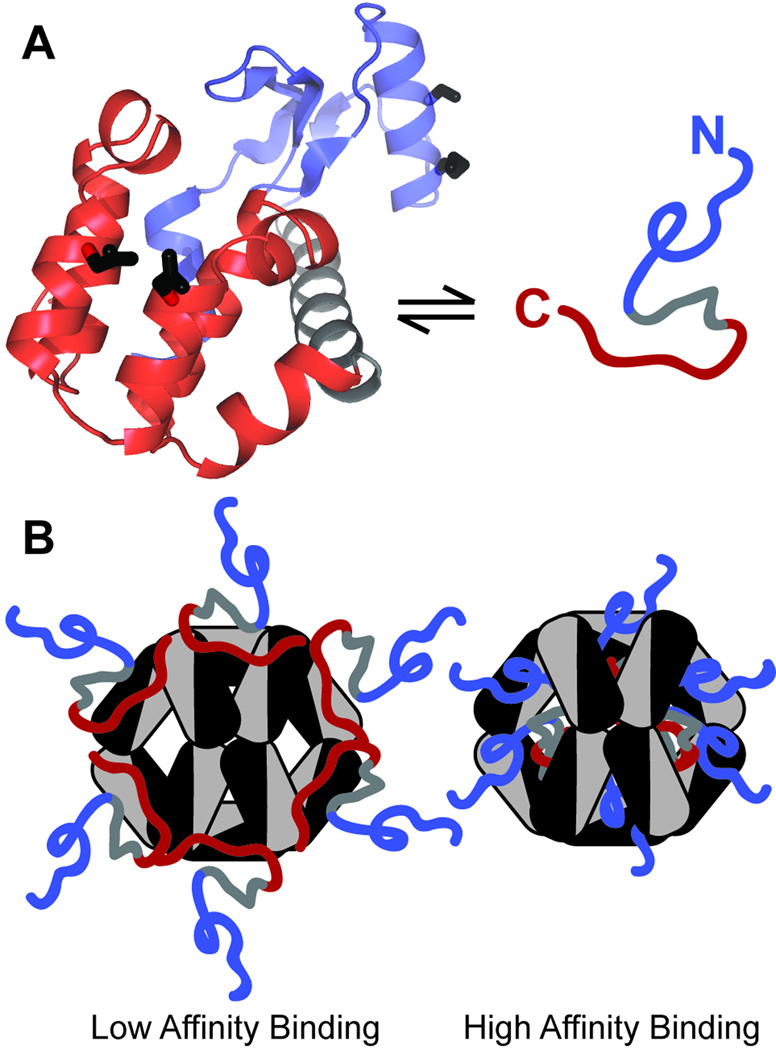
Small heat shock proteins (sHSP) make up a remarkably diverse group of molecular chaperones possessing a degree of structural plasticity unparalleled in other protein superfamilies. In the absence of chemical energy input, these stability sensors can sensitively recognize and bind destabilized proteins, even in the absence of gross misfolding. Cellular conditions regulate affinity toward client proteins, allowing tightly controlled switching and tuning of sHSP chaperone capacity. Perturbations of this regulation, through chemical modification or mutation, directly lead to a variety of disease states. This review explores the structural basis of sHSP oligomeric flexibility and the corresponding functional consequences in the context of a model describing sHSP activity with a set of three coupled thermodynamic equilibria. As current research illuminates many novel physiological roles for sHSP outside of their traditional duties as molecular chaperones, such a conceptual framework provides a sound foundation for describing these emerging functions in physiological and pathological processes.


Figures

Figure 1
Figure 1. Small heat shock protein architecture
(A) Schematic representation of Hsp16.5, Hsp16.9, and αA-crystallin drawn to scale depicting the α-crystallin domain (gray) flanked by N (blue) and C (red) terminal extensions with β strands as black arrows. (B) Quaternary structures of M. jannaschii Hsp 16.5 spherical 24-mer (left) and the dodecameric double disk of wheat Hsp16.9 (right) with N and C terminal extensions colored as (A) and the asymmetric α-crystallin domains of each dimer shaded in light and dark gray. (C) Hsp dimers with strands numbered as in (A). αA-Crystallin depicted as modeled from EPR distance analysis (55). Note the domain swapped strand 6 of Hsp16.5 and 16.9 and its absence in αA-crystallin.
Figure 2
Figure 2. CryoEM structural information for Hsp16.5 wild type and engineered variants
(A) Three-dimensional cryoEM reconstructions of WT Hsp16.5, Hsp16.5-TR, and Hsp16.5-P1 (left to right) shown cropped in half and radially color-coded (radius 10Å = blue; radius 90Å = red) with a 50 Å scale bar. (B) Three selected cryoEM class sum images of Hsp16.5-P1 displaying nearly perfect 2−, 3-, and 4-fold symmetry (left to right). (C) Three cryoEM class sum images of Hsp16.5-P1N selected to represent the diversity and lack of symmetry in the majority of the class sum images. Adapted with permission from (70). © 2009 The American Society for Biochemistry and Molecular Biology . All rights reserved.
Figure 3
Figure 3. CryoEM structure and pseudoatomic model of Hsp16.5-P1
(A) CryoEM structure at 10 Å resolution aligned along the 3- and 4-fold symmetry axes, left to right. The radial color coding is the same as in Figure 2. (B) Pseudoatomic model of Hsp16.5-P1 containing 48 α-crystallin domain monomers as viewed along the 3- and 4-fold symmetry axes. The independent monomer positions are colored either gold with magenta C terminal tails (chain A) or green with blue C terminal tails (chain B). (C) An α-crystallin domain dimer from the pseudoatomic model showing the two different C-terminal tail conformations (left). Superimposition of the two monomers within the dimer (right) shows the deviation in the angle of C-terminal tail relative to the α-crystallin domain. This angle deviation allows the dimer to accommodate packing around either the 3- or 4-fold openings of the assembly. Adapted with permission from (70). © 2009 The American Society for Biochemistry and Molecular Biology . All rights reserved.
Figure 4
Figure 4. Destabilized model substrate for Hsp binding analysis
(A) Structural superimposition of cysteineless pseudo wild type T4 Lysozyme (blue) and destabilized mutants L99A (green) and L46A (red) demonstrating preservation of tertiary structure. (B) Chemical denaturant unfolding curves of L99A and L46A as monitored by intrinsic tryptophan fluorescence depicting the relative destabilization of the two substrate mutations. (T4L WT ΔG=11.3 kcal/mol). (C) Binding isotherms demonstrating that the affinity of α-crystallin to destabilized T4L mutants is reflective of relative destabilization. Left shift of L99A curve demonstrates higher affinity binding. KD of both low and high affinity substrate binding modes are reported adjacent to features of the binding isotherm corresponding to each mode.
Figure 5
Figure 5. Structure of bound substrate and binding modes
(A) Crystal structure of T4L-L99A depicting representative residue pairs within the C terminal domain (red) labeled to probe the tertiary fold and in the N terminal region (blue) monitoring helical secondary structure proximities at i, i+4 residues shown in equilibrium with a schematic representation of an unfolded form. (B) Model depicting structural aspects of the low and high affinity binding modes. In each case there is extensive substrate unfolding with loss of proximities in labeled pairs tracking both secondary and tertiary structure. Labeled substrates report a net orientation with C termini located in a more conformationally restrictive, solvent inaccessible environment and N termini experiencing greater conformational mobility and solvent accessibility. The hydrodynamic radius of the high affinity binding mode is comparable to chaperone in absence of substrate and this radius increases in conditions favoring low affinity binding.
Figure 6
Figure 6. Hsp dissociation promotes activation and substrate binding
(A) Size exclusion chromatography elution profiles of wild type Hsp27 and a phosphorylation mimic in which the three serines phosphorylated by MAPK2 are replaced by aspartic acids to resemble this triply phosphorylated form (S15D/S78D/S82D). This phosphomimic shifts the oligomerization equilibrium in a manner favoring the complete disassembly of higher order oligomers observed in WT Hsp27 to smaller species. (B) The equation represents the dissociation of Hsp27 from a large oligomer (L) to a multimer (M) where p is an integer that accounts for the difference in the number of subunits between the two oligomeric states. Below, this equilibrium is depicted graphically in colors corresponding to the predominant SEC peaks in (A) as well as the binding isotherms in (C). (C) Binding is detected as quenching of a fluorescently labeled T4 lysozyme substrate. Phosphorylation induced dissociation of Hsp27-D3 drastically increases the affinity of Hsp27 for substrate compared to WT Hsp27. Combined with panel (A), this data reinforces a model of chaperone activation and regulation through phosphorylation induced changes in oligomeric state.
Scheme 1
Scheme 1
Coupled Equilibria Describing Minimalist Model

References

    1. Dill KA, Chan HS. From Levinthal to pathways to funnels. Nature Structural Biology. 1997;4:10–19. - PubMed
    1. Onuchic JN, Luthey-Schulten Z, Wolynes PG. Theory of protein folding: the energy landscape perspective. Annual Review of Physical Chemistry. 1997;48:545–600. - PubMed
    1. Dobson CM. Principles of protein folding, misfolding and aggregation. Seminars in Cell & Developmental Biology. 2004;15:3–16. - PubMed
    1. Bai Y, Sosnick TR, Mayne L, Englander SW. Protein folding intermediates: native-state hydrogen exchange. Science. 1995;269:192–197. - PMC - PubMed
    1. Englander SW, Mayne L, Krishna MMG. Protein folding and misfolding: mechanism and principles. Quarterly Reviews of Biophysics. 2007;40:287–326. - PMC - PubMed

Publication types