Coupled growth and division of model protocell membranes
- PMID: 19323552
- PMCID: PMC2669828
- DOI: 10.1021/ja900919c
Coupled growth and division of model protocell membranes
Abstract
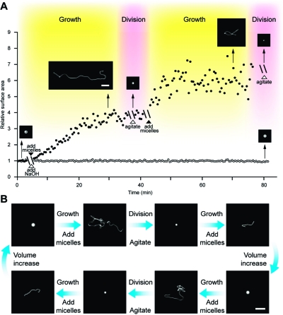
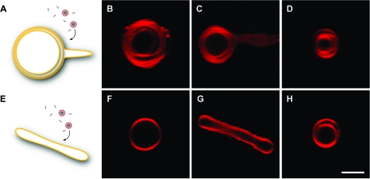
The generation of synthetic forms of cellular life requires solutions to the problem of how biological processes such as cyclic growth and division could emerge from purely physical and chemical systems. Small unilamellar fatty acid vesicles grow when fed with fatty acid micelles and can be forced to divide by extrusion, but this artificial division process results in significant loss of protocell contents during each division cycle. Here we describe a simple and efficient pathway for model protocell membrane growth and division. The growth of large multilamellar fatty acid vesicles fed with fatty acid micelles, in a solution where solute permeation across the membranes is slow, results in the transformation of initially spherical vesicles into long thread-like vesicles, a process driven by the transient imbalance between surface area and volume growth. Modest shear forces are then sufficient to cause the thread-like vesicles to divide into multiple daughter vesicles without loss of internal contents. In an environment of gentle shear, protocell growth and division are thus coupled processes. We show that model protocells can proceed through multiple cycles of reproduction. Encapsulated RNA molecules, representing a primitive genome, are distributed to the daughter vesicles. Our observations bring us closer to the laboratory synthesis of a complete protocell consisting of a self-replicating genome and a self-replicating membrane compartment. In addition, the robustness and simplicity of this pathway suggests that similar processes might have occurred under the prebiotic conditions of the early Earth.
Figures


Similar articles
-
Concentration-driven growth of model protocell membranes.J Am Chem Soc. 2012 Dec 26;134(51):20812-9. doi: 10.1021/ja310382d. Epub 2012 Dec 14. J Am Chem Soc. 2012. PMID: 23198690 Free PMC article.
-
Chemically Driven Division of Protocells by Membrane Budding.J Am Chem Soc. 2024 Dec 11;146(49):33359-33367. doi: 10.1021/jacs.4c08226. Epub 2024 Nov 27. J Am Chem Soc. 2024. PMID: 39603809 Free PMC article.
-
The origins of cellular life.Cold Spring Harb Perspect Biol. 2010 Sep;2(9):a002212. doi: 10.1101/cshperspect.a002212. Epub 2010 May 19. Cold Spring Harb Perspect Biol. 2010. PMID: 20484387 Free PMC article. Review.
-
Controlled growth of filamentous fatty acid vesicles under flow.Langmuir. 2014 Dec 16;30(49):14916-25. doi: 10.1021/la503933x. Epub 2014 Dec 1. Langmuir. 2014. PMID: 25402759 Free PMC article.
-
Early self-reproduction, the emergence of division mechanisms in protocells.Mol Biosyst. 2013 Feb 2;9(2):195-204. doi: 10.1039/c2mb25375e. Epub 2012 Dec 11. Mol Biosyst. 2013. PMID: 23232904 Review.
Cited by
-
L-form bacteria, cell walls and the origins of life.Open Biol. 2013 Jan 8;3(1):120143. doi: 10.1098/rsob.120143. Open Biol. 2013. PMID: 23303308 Free PMC article. Review.
-
Growth, replication and division enable evolution of coacervate protocells.Chem Commun (Camb). 2022 Oct 6;58(80):11183-11200. doi: 10.1039/d2cc03541c. Chem Commun (Camb). 2022. PMID: 36128910 Free PMC article. Review.
-
Fast bilayer-micelle fusion mediated by hydrophobic dipeptides.Biophys J. 2021 Jun 1;120(11):2330-2342. doi: 10.1016/j.bpj.2021.04.012. Epub 2021 Apr 19. Biophys J. 2021. PMID: 33887225 Free PMC article.
-
Mechanomicrobiology: how bacteria sense and respond to forces.Nat Rev Microbiol. 2020 Apr;18(4):227-240. doi: 10.1038/s41579-019-0314-2. Epub 2020 Jan 20. Nat Rev Microbiol. 2020. PMID: 31959911 Review.
-
Passive endocytosis in model protocells.Proc Natl Acad Sci U S A. 2023 Jun 13;120(24):e2221064120. doi: 10.1073/pnas.2221064120. Epub 2023 Jun 5. Proc Natl Acad Sci U S A. 2023. PMID: 37276401 Free PMC article.
References
Publication types
MeSH terms
Substances
Grants and funding
LinkOut - more resources
Full Text Sources
Molecular Biology Databases