

Development of an ultraperformance liquid chromatography/mass spectrometry method to quantify cisplatin 1,2 intrastrand guanine-guanine adducts
- PMID: 19323581
- PMCID: PMC2728554
- DOI: 10.1021/tx800481j
Development of an ultraperformance liquid chromatography/mass spectrometry method to quantify cisplatin 1,2 intrastrand guanine-guanine adducts
Abstract
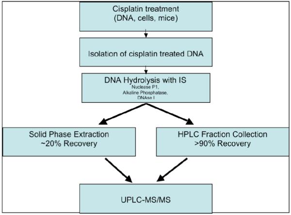
Platinum chemotherapeutic agents have been widely used in the treatment of cancer. Cisplatin was the first of the platinum-based chemotherapeutic agents and therefore has been extensively studied as an antitumor agent since the late 1960s. Because this agent forms several DNA adducts, a highly sensitive and specific quantitative assay is needed to correlate the molecular dose of individual adducts with the effects of treatment. An ultraperformance liquid chromatography tandem mass spectrometry (UPLC-MS/MS) assay for quantification of 1,2 guanine-guanine intrastrand cisplatin adducts [CP-d(GpG)], using (15)N(10) CP-d(GpG) as an internal standard, was developed. The internal standard was characterized by MS/MS, and its concentration was validated by inductively coupled plasma mass spectrometry. Samples containing CP-d(GpG) in DNA were purified by enzyme hydrolysis, centrifugal filtration, and HPLC with fraction collection prior to quantification by UPLC-MS/MS in the selective reaction monitoring mode [m/z 412.5-->248.1 for CP-d(GpG); m/z 417.5-->253.1 for [(15)N(10)] CP-d(GpG)]. The recovery of standards was >90%, and quantification was unaffected by increasing concentrations of calf thymus DNA. This method utilizes 25 mug of DNA per injection. The limit of quantification was 3 fmol or 3.7 adducts per 10(8) nucleotides, which approaches the sensitivity of the (32)P postlabeling method for this adduct. These data suggested that this method is suitable for in vitro and in vivo assessment of CP-d(GpG) adducts formed by cisplatin and carboplatin. Subsequently, the method was applied to studies using ovarian carcinoma cell lines and C57/BL6 mice to illustrate that this method is capable of quantifying CP-d(GpG) adducts using biologically relevant systems and doses. The development of biomarkers to determine tissue-specific molecular dosimetry during treatment will lead to a more complete understanding of both therapeutic and adverse effects of cisplatin and carboplatin. This will support the refinement of therapeutic regimes and appropriate individualized treatment protocols.
Figures


Comment in
-
Response to Interpretation of Mass Spectral Data for the Cisplatin 1,2 Intrastrand Guanine-Guanine Adduct.Chem Res Toxicol. 2018 Nov 19;31(11):1108. doi: 10.1021/acs.chemrestox.8b00260. Epub 2018 Oct 16. Chem Res Toxicol. 2018. PMID: 30215251 No abstract available.
-
Interpretation of Mass Spectral Data for the Cisplatin 1,2 Intrastrand Guanine-Guanine Adduct.Chem Res Toxicol. 2018 Nov 19;31(11):1106-1107. doi: 10.1021/acs.chemrestox.8b00134. Epub 2018 Oct 16. Chem Res Toxicol. 2018. PMID: 30280575 Free PMC article. No abstract available.
Similar articles
-
Debio 0507 primarily forms diaminocyclohexane-Pt-d(GpG) and -d(ApG) DNA adducts in HCT116 cells.Cancer Chemother Pharmacol. 2012 Mar;69(3):665-77. doi: 10.1007/s00280-011-1744-3. Epub 2011 Oct 4. Cancer Chemother Pharmacol. 2012. PMID: 21968950 Free PMC article.
-
Determination of cisplatin 1,2-intrastrand guanine-guanine DNA adducts in human leukocytes by high-performance liquid chromatography coupled to inductively coupled plasma mass spectrometry.Chem Res Toxicol. 2010 Aug 16;23(8):1313-21. doi: 10.1021/tx100023c. Chem Res Toxicol. 2010. PMID: 20666396
-
An approach based on ultra-high pressure liquid chromatography-tandem mass spectrometry to quantify O6-methyl and O6-carboxymethylguanine DNA adducts in intestinal cell lines.J Chromatogr A. 2012 Sep 28;1257:25-33. doi: 10.1016/j.chroma.2012.07.040. Epub 2012 Jul 20. J Chromatogr A. 2012. PMID: 22921361
-
Quantitative high-performance liquid chromatography-electrospray ionization-tandem mass spectrometry analysis of the adenine-guanine cross-links of 1,2,3,4-diepoxybutane in tissues of butadiene-exposed B6C3F1 mice.Chem Res Toxicol. 2008 May;21(5):1163-70. doi: 10.1021/tx800051y. Epub 2008 Apr 29. Chem Res Toxicol. 2008. PMID: 18442269 Free PMC article.
-
A rare example of three abundant conformers in one retro model of the cisplatin-DNA d(GpG) intrastrand cross link. Unambiguous evidence that guanine O6 to carrier amine ligand hydrogen bonding is not important. possible effect of the Lippard base pair step adjacent to the lesion on carrier ligand hydrogen bonding in DNA adducts.J Am Chem Soc. 2001 Sep 26;123(38):9345-55. doi: 10.1021/ja010483m. J Am Chem Soc. 2001. PMID: 11562217
Cited by
-
Nuclease digestion and mass spectrometric characterization of oligodeoxyribonucleotides containing 1,2-GpG, 1,2-ApG, and 1,3-GpXpG cisplatin intrastrand cross-links.Clin Chim Acta. 2013 May;420:160-70. doi: 10.1016/j.cca.2012.12.010. Epub 2012 Dec 22. Clin Chim Acta. 2013. PMID: 23266768 Free PMC article.
-
Interpretation of Mass Spectral Data for the Cisplatin 1,2 Intrastrand Guanine-Guanine Adduct.Chem Res Toxicol. 2018 Nov 19;31(11):1106-1107. doi: 10.1021/acs.chemrestox.8b00134. Epub 2018 Oct 16. Chem Res Toxicol. 2018. PMID: 30280575 Free PMC article. No abstract available.
-
Mitochondrial Dysfunction in Chemotherapy-Induced Peripheral Neuropathy (CIPN).Toxics. 2015 Jun 5;3(2):198-223. doi: 10.3390/toxics3020198. Toxics. 2015. PMID: 29056658 Free PMC article. Review.
-
Debio 0507 primarily forms diaminocyclohexane-Pt-d(GpG) and -d(ApG) DNA adducts in HCT116 cells.Cancer Chemother Pharmacol. 2012 Mar;69(3):665-77. doi: 10.1007/s00280-011-1744-3. Epub 2011 Oct 4. Cancer Chemother Pharmacol. 2012. PMID: 21968950 Free PMC article.
-
Approaching Sites of Action of Temozolomide for Pharmacological and Clinical Studies in Glioblastoma.Biomedicines. 2021 Dec 21;10(1):1. doi: 10.3390/biomedicines10010001. Biomedicines. 2021. PMID: 35052681 Free PMC article.
References
-
- Reardon JT, Vaisman A, Chaney SG, Sancar A. Efficient nucleotide excision repair of cisplatin, oxaliplatin, and Bis-aceto-ammine-dichloro-cyclohexylamine-platinum(IV) (JM216) platinum intrastrand DNA diadducts. Cancer Res. 1999;59:3968–3971. - PubMed
-
- Zamble DB, Mu D, Reardon JT, Sancar A, Lippard SJ. Repair of cisplatin--DNA adducts by the mammalian excision nuclease. Biochemistry. 1996;35:10004–10013. - PubMed
-
- Eastman A. The formation, isolation and characterization of DNA adducts produced by anticancer platinum complexes. Pharmacol. Ther. 1987;34:155–166. - PubMed
Publication types
MeSH terms
Substances
Grants and funding
LinkOut - more resources
Full Text Sources
Other Literature Sources
Miscellaneous
