

Alteration in functional brain systems after electrical injury
- PMID: 19323610
- PMCID: PMC6468944
- DOI: 10.1089/neu.2008.0867
Alteration in functional brain systems after electrical injury
Abstract
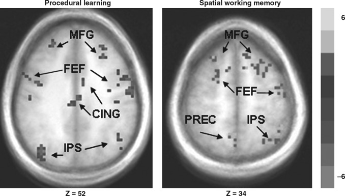
Neuropsychological studies in electrical injury patients have reported deficits in attention, learning, and working memory, but the neural substrates of these deficits remain poorly characterized. In this study we sought to examine whether electrical injury subjects demonstrate abnormal patterns of brain activation during working memory and procedural learning tasks. Fourteen electrical injury subjects and fifteen demographically matched healthy control subjects performed a spatial working memory paradigm and a procedural learning paradigm during functional MRI studies. For the spatial working memory task, electrical injury patients exhibited significantly greater activation in the middle frontal gyrus and motor and posterior cingulate cortices. Increased activation in EI subjects also was observed on a visually-guided saccade task in several sensorimotor regions, including the frontal and parietal eye fields and striatum. On the procedural learning task, electrical injury patients exhibited significantly less activation in the middle frontal gyrus, anterior cingulate cortex, and frontal eye fields than controls. This is the first study to document task-dependent, system-level cortical and subcortical dysfunction in individuals who had experienced an electrical shock trauma.
Figures
References
-
- Beck A. Steer R. Brown G. Beck Depression Inventory–II. The Psychological Corporation; San Antonio, TX: 1987.
-
- Conner C.K. Manual for the Conner's Continuous Performance Test–II. Multi health Systems; Tanawanda, NY: 2002.
-
- Cox R.W. AFNI: Software for analysis and visualization of functional magnetic neuroimages. Comput. Biomed. Res. 1996;29:162–173. - PubMed
-
- Delis D.C. Kramer J.H. Kaplan E. Ober B.A. California Verbal Learning Test, Second Adult Version Manual. The Psychological Corporation; San Antonio, TX: 2000.
MeSH terms
Grants and funding
LinkOut - more resources
Full Text Sources
Medical
