

Ultrasound in evaluation of post-interventional femoral vein obstruction: a case report
- PMID: 19323809
- PMCID: PMC2667400
- DOI: 10.1186/1476-7120-7-14
Ultrasound in evaluation of post-interventional femoral vein obstruction: a case report
Abstract
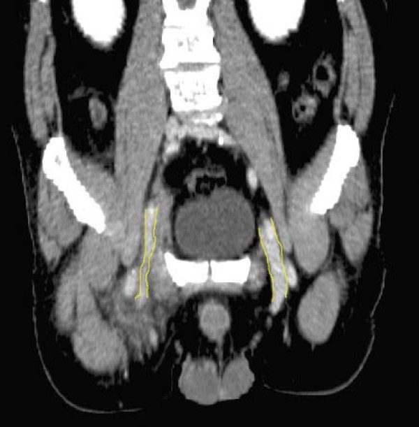
Ultrasound is the preferred imaging modality in diagnosis of vascular complications following cardiac catheterization and intervention. In some cases, however, bleeding surrounding the femoral vessels, may severely distort the color Doppler images, making detection of venous complications especially difficult. This report refers to such a case where post-catheterization haematoma was suspected to cause an obstruction of the femoral vein. Spectral Doppler recordings of blood flow in the common femoral vein, up-stream, distal to the hemorrhagic area, confirmed the diagnosis of obstruction by demonstrating changes in the venous flow pattern in the common femoral vein, consistent with venous hypertension. Due to the poor quality of the ultrasound images, the exact cause of the obstruction had to be established by another imaging modality, not affected by haemorrhages. CT showed that the common femoral vein was compressed at the puncture site by surrounding haemorrhages. Thus, when bleeding due to cardiac catheterization is associated with possible venous obstruction and findings by color Doppler are equivocal due to degradation of the color-Doppler image, detection of venous hypertension by spectral Doppler, performed distal to the bleeding area, strongly supports the presence of venous obstruction where the exact cause may be established by CT.
Figures


Similar articles
-
[Color-coded Doppler in the diagnosis of vascular complications following heart catheterization].Rev Esp Cardiol. 1992 Jun-Jul;45(6):374-80. Rev Esp Cardiol. 1992. PMID: 1631385 Spanish.
-
The safety and efficacy of the StarClose Vascular Closure System: the ultrasound substudy of the CLIP study.Catheter Cardiovasc Interv. 2006 Nov;68(5):684-9. doi: 10.1002/ccd.20898. Catheter Cardiovasc Interv. 2006. PMID: 17039509 Clinical Trial.
-
Post-cardiac catheterization femoral fistula corrected by ultrasound-guided compression.JBR-BTR. 2005 Jan-Feb;88(1):7-11. JBR-BTR. 2005. PMID: 15792161
-
[Color Doppler evaluation and diagnosis of local complications after arterial endovascular procedures].Recenti Prog Med. 2012 Sep;103(9):337-47. doi: 10.1701/1136.12528. Recenti Prog Med. 2012. PMID: 23023022 Review. Italian.
-
A Synovial Cyst Originating from the Hip Joint as a Rare Cause of Recurrent Femoral Vein Thrombosis: Case Report and Literature Review.Ann Vasc Surg. 2017 Aug;43:313.e13-313.e15. doi: 10.1016/j.avsg.2017.02.016. Epub 2017 May 4. Ann Vasc Surg. 2017. PMID: 28479436 Review.
References
-
- Messina LM, Brothers TE, Wakefield TW, Zelenock GB, Lindenauer SM, Greenfield LJ, et al. Clinical characteristics and surgical management of vascular complications in patients undergoing cardiac catheterization: interventional versus diagnostic procedures. J Vasc Surg. 1991;13:593–600. doi: 10.1067/mva.1991.27611. - DOI - PubMed
-
- Bach AM, Hann LE. When the common femoral vein is revealed as flattened on spectral Doppler sonography: is it a reliable sign for diagnosis of proximal venous obstruction? AJR Am J Roentgenol. 1997;168:733–736. - PubMed
Publication types
MeSH terms
LinkOut - more resources
Full Text Sources
