. 2009 Mar 31;53(13):1138-45.
doi: 10.1016/j.jacc.2008.11.052.

Delayed-enhanced magnetic resonance imaging in nonischemic cardiomyopathy: utility for identifying the ventricular arrhythmia substrate

Affiliations

Delayed-enhanced magnetic resonance imaging in nonischemic cardiomyopathy: utility for identifying the ventricular arrhythmia substrate

Frank M Bogun et al. J Am Coll Cardiol. .

Abstract

Objectives: The purpose of this study was to assess the value of delayed-enhanced magnetic resonance imaging (DE-MRI) to guide ablation of ventricular arrhythmias in patients with nonischemic cardiomyopathy (NIC).

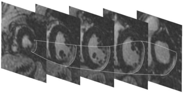
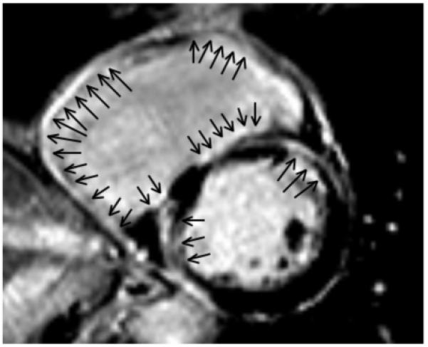
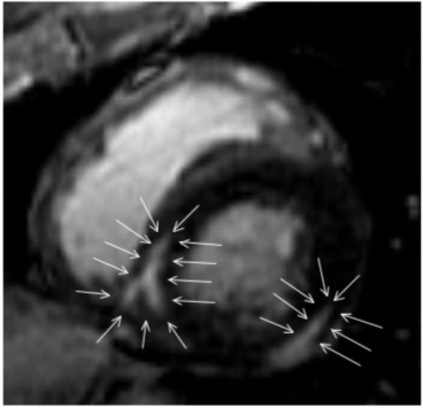
Background: In patients with NIC, ventricular arrhythmias often are associated with scar tissue. DE-MRI can be used to precisely define scar tissue.

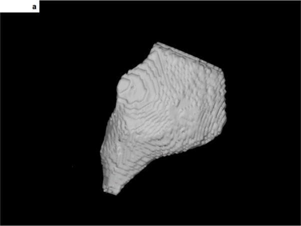
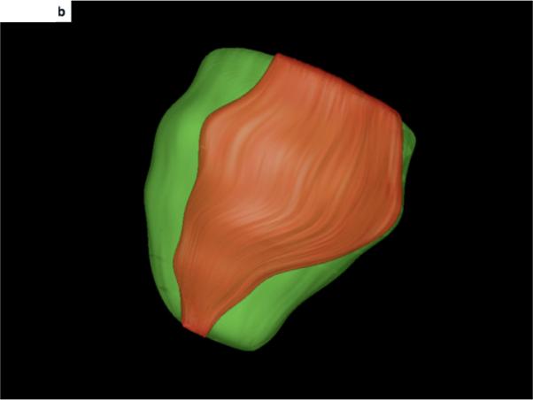
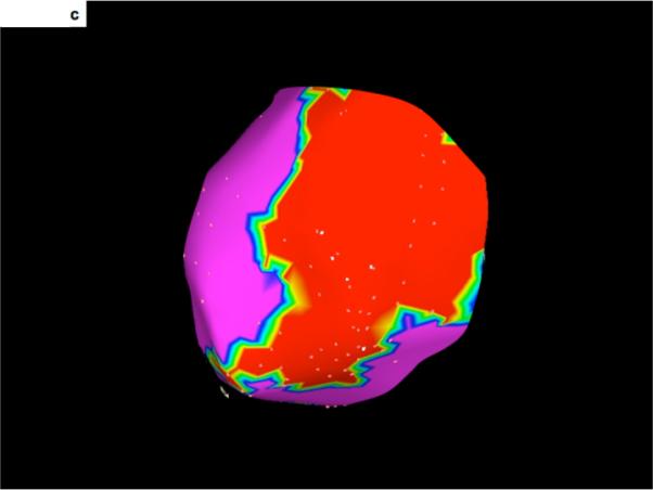
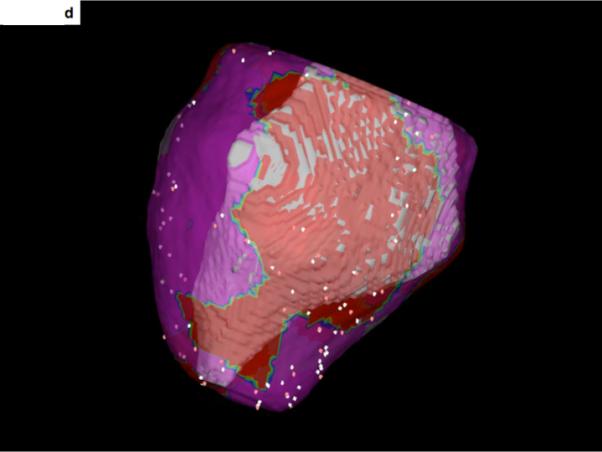
Methods: DE-MRI was performed in 29 consecutive patients (mean age 50 +/- 15 years) with NIC (mean ejection fraction 37 +/- 9%) referred for catheter ablation of ventricular tachycardia (VT) or premature ventricular complexes (PVCs). Scar was extracted from DE-MRIs and was then integrated into the electroanatomic map. Mapping data were correlated with respect to the localization of scar tissue.

Results: Scar was identified by DE-MRI in 14 of 29 patients. Nine of these patients had VT and 5 had PVCs. In 5 of the patients there was predominantly endocardial scar, and mapping and ablation of arrhythmias was effectively performed from the endocardium in all 5 patients. In 2 patients scar was either intramural or epicardial with extension to the endocardium. In both patients with partial endocardial scar extension, the ablation was effective in eliminating some but not all arrhythmias. In 2 patients most of the scar tissue was confined to the epicardium; mapping identified and eliminated an epicardial origin in both patients. No effect on arrhythmias could be achieved in the other 5 patients with predominantly intramural scar.

Conclusions: DE-MRI in patients without prior infarctions can help to identify the arrhythmogenic substrate; furthermore, it helps to plan an appropriate mapping and ablation strategy.


Figures

Figure 1
Figure 1. Stack of short axis DE-MRI pictures showing delayed enhancement in the epicardial left ventricular lateral wall
Although the free wall is thinned, the endocardium is relatively preserved. The epicardial contours of the scar are traced in dotted lines. An endocardial radiofrequency catheter ablation procedure for VT was unsuccessful. Epicardial ablation successfully eliminated all 3 inducible VTs.
Figure 2
Figure 2. 3-D display and integration of scar within the electroanatomic map
A- The 3-dimensional display of scar tissue (grey) that was extracted from the DE-MRI shown in Figure 1. The inferolateral aspect of the epicardial scar is shown. B- The 3-dimensional display of scar (orange) and the surrounding epicardium (green) of the left ventricle. The heart is oriented in the same manner as in Figure 2A, showing the inferolateral aspect of the scar extending from the left ventricular base to the apex. C- An epicardial voltage map. Low voltage areas (<1.5mV) are displayed in red). The scar shown in Figure 2A and 2B corresponds with the low voltage area shown here. D- Merging of the voltage map with the 3-dimensional reconstruction of the scar seen on DE-MRI. The region of low voltage corresponds to the location of the DE-MRI scar.
Figure 3
Figure 3. A short-axis view of the mid-portion of the right and left ventricles
The scar involves predominantly the right ventricular endocardium. The right ventricle shows enodcardial delayed enhancement (arrow) that is transmural in the right ventricular free wall and mostly endocardial at the right ventricular septum. Endocardial scar also is present in the left ventricle, with some transmural components. All 4 inducible VTs were ablated at the right ventricular endocardium in this patient.
Figure 4
Figure 4. A short-axis view of the mid-portion of the left ventricle
There is delayed enhancement that is predominantly mid-myocardial at the inferior free wall. The scar is intramural (arrows) and extends to the epicardium, with sparing of the endocardium. The scar located at the postero-septal wall is completely intramural (arrows). Endocardial radiofrequency catheter ablation was unsuccessful in this patient. Limited epicardial mapping from within the coronary sinus did not show early activation during the induced VTs, and mapping was not performed within the pericardial space.

Comment in

References

    1. Nazarian S, Bluemke DA, Lardo AC, Zviman MM, Watkins SP, Dickfeld TL, Meininger GR, Roguin A, Calkins H, Tomaselli GF, Weiss RG, Berger RD, Lima JA, Halperin HR. Magnetic resonance assessment of the substrate for inducible ventricular tachycardia in nonischemic cardiomyopathy. Circulation. 2005;112:2821–5. - PMC - PubMed
    1. Assomull RG, Prasad SK, Lyne J, Smith G, Burman ED, Khan M, Sheppard MN, Poole-Wilson PA, Pennell DJ. Cardiovascular magnetic resonance, fibrosis, and prognosis in dilated cardiomyopathy. J Am Coll Cardiol. 2006;48:1977–85. - PubMed
    1. Wu KC, Weiss RG, Thiemann DR, Kitagawa K, Schmidt A, Dalal D, Lai S, Bluemke DA, Gerstenblith G, Marban E, Tomaselli GF, Lima JA. Late gadolinium enhancement by cardiovascular magnetic resonance heralds an adverse prognosis in nonischemic cardiomyopathy. J Am Coll Cardiol. 2008;51:2414–21. - PMC - PubMed
    1. McCrohon JA, Moon JC, Prasad SK, McKenna WJ, Lorenz CH, Coats AJ, Pennell DJ. Differentiation of heart failure related to dilated cardiomyopathy and coronary artery disease using gadolinium-enhanced cardiovascular magnetic resonance. Circulation. 2003;108:54–9. - PubMed
    1. De Cobelli F, Pieroni M, Esposito A, Chimenti C, Belloni E, Mellone R, Canu T, Perseghin G, Gaudio C, Maseri A, Frustaci A, Del Maschio A. Delayed gadolinium-enhanced cardiac magnetic resonance in patients with chronic myocarditis presenting with heart failure or recurrent arrhythmias. J Am Coll Cardiol. 2006;47:1649–54. - PubMed