

Manual MRI parcellation of the frontal lobe
- PMID: 19324532
- PMCID: PMC2728025
- DOI: 10.1016/j.pscychresns.2009.01.006
Manual MRI parcellation of the frontal lobe
Abstract
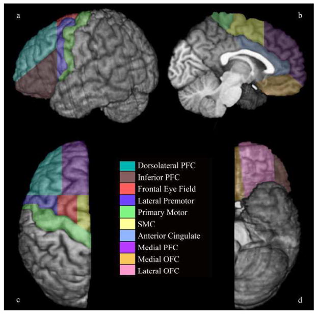
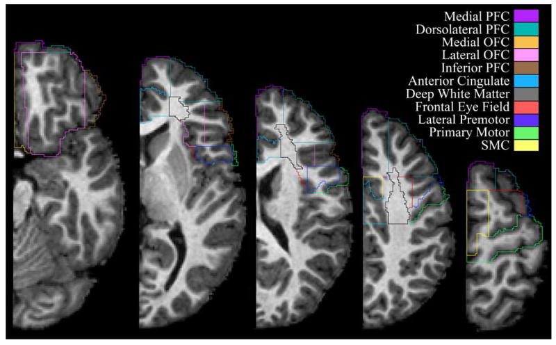
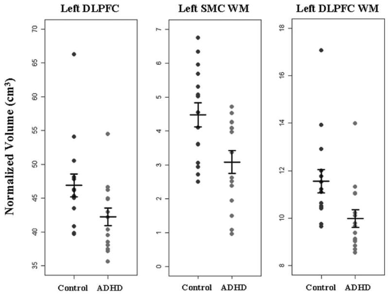
The ability to examine associations between neuropsychiatric conditions and functionally relevant frontal lobe sub-regions is a fundamental goal in neuropsychiatry, but methods for identifying frontal sub-regions in MR (magnetic resonance) images are not well established. Prior published techniques have principally defined gyral regions that do not necessarily correspond to known functional divisions. We present a method in which sulcal-gyral landmarks are used to manually delimit functionally relevant regions within the frontal lobe: primary motor cortex, anterior cingulate, deep white matter, premotor cortex regions (supplementary motor complex (SMC), frontal eye field and lateral premotor cortex) and prefrontal cortex (PFC) regions (medial PFC, dorsolateral PFC (DLPFC), inferior PFC, lateral orbitofrontal cortex (OFC) and medial OFC). Feasibility was tested by applying the protocol to brain MR data from 15 boys with attention-deficit/hyperactivity disorder (ADHD) and 15 typically developing controls, 8-12 years old. Intra- and inter-rater intraclass correlation coefficients were calculated using parcellation volumes from a subset of that group. Inter-rater results for the 22 hemisphere specific sub-regions ranged from 0.724 to 0.997, with all but seven values above 0.9. Boys with ADHD showed significantly smaller left hemisphere SMC and DLPFC volumes after normalization for total cerebral volume. These findings support the method as a reliable and valid technique for parcellating the frontal lobe into functionally relevant sub-regions.
Figures
Similar articles
-
Comprehensive examination of frontal regions in boys and girls with attention-deficit/hyperactivity disorder.J Int Neuropsychol Soc. 2011 Nov;17(6):1047-57. doi: 10.1017/S1355617711001056. Epub 2011 Sep 19. J Int Neuropsychol Soc. 2011. PMID: 21923979 Free PMC article.
-
Automated MRI parcellation of the frontal lobe.Hum Brain Mapp. 2014 May;35(5):2009-26. doi: 10.1002/hbm.22309. Epub 2013 Jul 29. Hum Brain Mapp. 2014. PMID: 23897577 Free PMC article.
-
MRI parcellation of the frontal lobe in boys with attention deficit hyperactivity disorder or Tourette syndrome.Psychiatry Res. 2002 Nov 30;116(1-2):63-81. doi: 10.1016/s0925-4927(02)00066-5. Psychiatry Res. 2002. PMID: 12426035
-
[Structural and functional neuroanatomy of attention-deficit hyperactivity disorder (ADHD)].Encephale. 2009 Apr;35(2):107-14. doi: 10.1016/j.encep.2008.01.005. Epub 2008 Jul 7. Encephale. 2009. PMID: 19393378 Review. French.
-
Cortical and Subcortical Gray Matter Volume in Youths With Conduct Problems: A Meta-analysis.JAMA Psychiatry. 2016 Jan;73(1):64-72. doi: 10.1001/jamapsychiatry.2015.2423. JAMA Psychiatry. 2016. PMID: 26650724 Review.
Cited by
-
Sex-Based Dissociation of White Matter Microstructure in Children With Attention-Deficit/Hyperactivity Disorder.J Am Acad Child Adolesc Psychiatry. 2015 Nov;54(11):938-46. doi: 10.1016/j.jaac.2015.08.014. Epub 2015 Sep 5. J Am Acad Child Adolesc Psychiatry. 2015. PMID: 26506584 Free PMC article.
-
Comprehensive examination of frontal regions in boys and girls with attention-deficit/hyperactivity disorder.J Int Neuropsychol Soc. 2011 Nov;17(6):1047-57. doi: 10.1017/S1355617711001056. Epub 2011 Sep 19. J Int Neuropsychol Soc. 2011. PMID: 21923979 Free PMC article.
-
Automated MRI parcellation of the frontal lobe.Hum Brain Mapp. 2014 May;35(5):2009-26. doi: 10.1002/hbm.22309. Epub 2013 Jul 29. Hum Brain Mapp. 2014. PMID: 23897577 Free PMC article.
-
Development of a methodology for the volume estimation of the prefrontal cortical subfields in very pre-term infants using magnetic resonance imaging and stereology.Asian Biomed (Res Rev News). 2025 Sep 2;19(4):174-182. doi: 10.2478/abm-2025-0023. eCollection 2025 Aug. Asian Biomed (Res Rev News). 2025. PMID: 40904614 Free PMC article.
-
Distinct frontal lobe morphology in girls and boys with ADHD.Neuroimage Clin. 2014 Dec 10;7:222-9. doi: 10.1016/j.nicl.2014.12.010. eCollection 2015. Neuroimage Clin. 2014. PMID: 25610784 Free PMC article.
References
-
- Acosta MT, Pearl PL. Imaging data in autism: from structure to malfunction. Seminars in Pediatric Neurology. 2004;11:205–213. - PubMed
-
- Alvarez JA, Emory E. Executive Function and the Frontal Lobes: A Meta-Analytic Review. Neuropsychology Review 2006 - PubMed
-
- Ashe J, Lungu OV, Basford AT, Lu X. Cortical control of motor sequences. Current Opinion in Neurobiology. 2006;16:213–221. - PubMed
-
- Aylward EH, Augustine A, Li Q, Barta PE, Pearlson GD. Measurement of frontal lobe volume on magnetic resonance imaging scans. Psychiatry Research. 1997;75:23–30. - PubMed
-
- Braver TS, Cohen JD, Nystrom LE, Jonides J, Smith E, Noll DC. A parametric study of prefrontal cortex involvement in human working memory. Neuroimage. 1997;5:49–62. - PubMed
Publication types
MeSH terms
Grants and funding
LinkOut - more resources
Full Text Sources
Medical
Miscellaneous
