. 2009 Jun 5;284(23):15835-46.
doi: 10.1074/jbc.M807846200. Epub 2009 Mar 25.

Mechanisms of base selection by human single-stranded selective monofunctional uracil-DNA glycosylase

Affiliations

Mechanisms of base selection by human single-stranded selective monofunctional uracil-DNA glycosylase

Agus Darwanto et al. J Biol Chem. .

Abstract

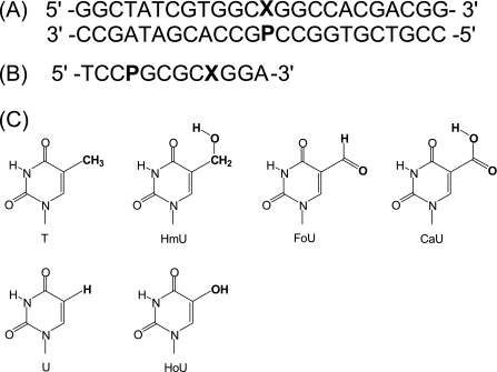
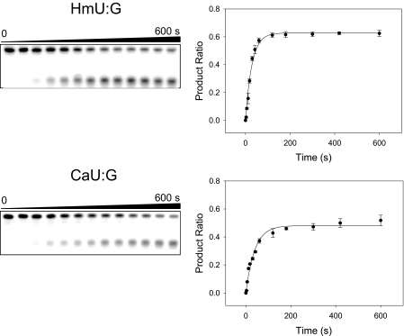
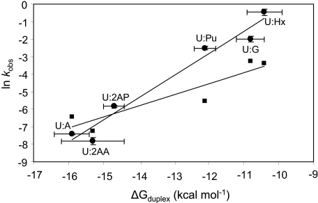
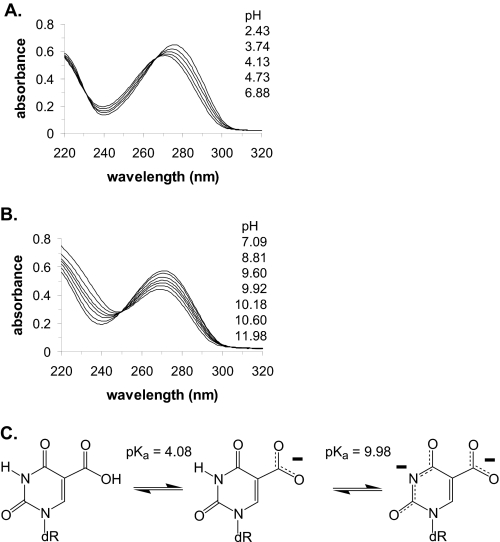
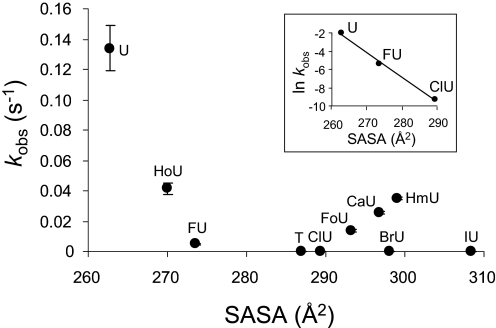
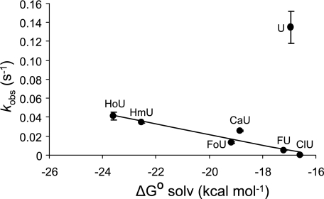
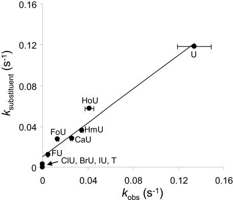
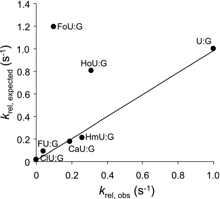
hSMUG1 (human single-stranded selective monofunctional uracil-DNA glyscosylase) is one of three glycosylases encoded within a small region of human chromosome 12. Those three glycosylases, UNG (uracil-DNA glycosylase), TDG (thymine-DNA glyscosylase), and hSMUG1, have in common the capacity to remove uracil from DNA. However, these glycosylases also repair other lesions and have distinct substrate preferences, indicating that they have potentially redundant but not overlapping physiological roles. The mechanisms by which these glycosylases locate and selectively remove target lesions are not well understood. In addition to uracil, hSMUG1 has been shown to remove some oxidized pyrimidines, suggesting a role in the repair of DNA oxidation damage. In this paper, we describe experiments in which a series of oligonucleotides containing purine and pyrimidine analogs have been used to probe mechanisms by which hSMUG1 distinguishes potential substrates. Our results indicate that the preference of hSMUG1 for mispaired uracil over uracil paired with adenine is best explained by the reduced stability of a duplex containing a mispair, consistent with previous reports with Escherichia coli mispaired uracil-DNA glycosylase. We have also extended the substrate range of hSMUG1 to include 5-carboxyuracil, the last in the series of damage products from thymine methyl group oxidation. The properties used by hSMUG1 to select damaged pyrimidines include the size and free energy of solvation of the 5-substituent but not electronic inductive properties. The observed distinct mechanisms of base selection demonstrated for members of the uracil glycosylase family help explain how considerable diversity in chemical lesion repair can be achieved.


Figures

FIGURE 1.
FIGURE 1.
Sequences of oligonucleotides and structures of uracil analogs used in this study. A, oligonucleotide duplex used for glycosylase assays, in which X represents thymine, uracil, or a 5-substituted uracil analog and P is a purine. B, sequence of the self-complementary oligonucleotide used for the determination of duplex stability. C, structures of uracil analogs.
FIGURE 2.
FIGURE 2.
Kinetic study of hSMUG1 cleavage of 5-substituted uracil analogues paired with guanine illustrating the gel electrophoretic assay (left) and time-dependent product ratios (right). Single-turnover reactions were performed at 37 °C with 50 nm substrate and 200 nm hSMUG1 in the reaction buffer, as described under “Materials and Methods.” Top, HmU:G as a substrate; bottom, CaU:G as a substrate.
FIGURE 3.
FIGURE 3.
Linear relationship between the free energy of duplex formation, ΔGduplex, and observed cleavage rate constants. MUG data from Ref. is presented as squares, and hSMUG1 from this study is presented as circles. The natural logarithms of the observed rate constants (ln kobs) for MUG and hSMUG1 are plotted versus ΔGduplex. Straight lines are obtained for hSMUG1 with a slope of 1.26, intercept of 12.35, and r2 = 0.96 and for MUG with a slope of 0.63, intercept of 3.02, and r2 = 0.85.
FIGURE 4.
FIGURE 4.
Ionization of 5-carboxyuracil. A, ionization of the 5-carboxyl group with a pKa of 4.08. B, ionization of the N3 ring nitrogen with a pKa of 9.98. C, structures of the neutral and ionized forms of CaU.
FIGURE 5.
FIGURE 5.
Relationship between SASA and glycosylase kinetics. The observed rate constants (kobs) for hSMUG1 cleavage of uracil analogs paired opposite guanine are plotted against the SASA. Inset, the impact of the size alone on the apparent rate constant. In the inset, values of ln kobs for U:G, FU:G, and ClU:G are plotted versus SASA. A straight line is obtained, with slope of −0.27, intercept of 69.19, and r2 = 0.99.
FIGURE 6.
FIGURE 6.
Relationship between glycosylase cleavage rates and ΔG0 solvation. The observed rate constants (kobs) for hSMUG1 cleavage of uracil analogs opposite guanine are plotted against ΔG0 solvation. A line is drawn through all the points except uracil. The slope of this line is −5.58 × 10⁻³ with intercept of −8.96 × 10⁻², and r2 is 0.90.
FIGURE 7.
FIGURE 7.
The comparison of observed rate constants (kobs) and expected rate constants based upon substituent characteristics (ksubstituent). The observed rate is plotted against the expected rate based upon Equation 11. A straight line is obtained, with slope of 0.83, intercept of 0.01, and r2 = 0.97.
FIGURE 8.
FIGURE 8.
Relationship between observed relative rate constants (krel,obs) and expected relative rate constants based on duplex formation energy of the oligonucleotide, size, and free energy of solvation of the uracil 5-substituent (krel,expected). The expected relative rates are calculated with respect to the rate of cleavage of the U:G base pair according to Equation 13. A straight line is obtained for several of the analogs with slope of 0.98, intercept of 9.20 × 10⁻³, and r2 = 0.99. Values for the FoU:G and HoU:G pairs do not fall on this line.

Similar articles

Cited by

References

    1. Sousa M. M., Krokan H. E., Slupphaug G. ( 2007) Mol. Aspects Med. 28, 276– 306 - PubMed
    1. Slupphaug G., Eftedal I., Kavli B., Bharati S., Helle N. M., Haug T., Levine D. W., Krokan H. E. ( 1995) Biochemistry 34, 128– 138 - PubMed
    1. Neddermann P., Gallinari P., Lettieri T., Schmid D., Truong O., Hsuan J. J., Weibauer K., Jiricny J. ( 1996) J. Biol. Chem. 217, 12767– 12774 - PubMed
    1. Nilsen H., Haushalter K. A., Robins P., Barnes D. E., Verdine G. L., Lindahl T. ( 2001) EMBO J. 20, 4278– 4286 - PMC - PubMed
    1. Pearl L. H. ( 2000) Mutat. Res. 460, 165– 181 - PubMed

Publication types

LinkOut - more resources