. 2009 May 15;284(20):13348-13354.
doi: 10.1074/jbc.M809449200. Epub 2009 Mar 26.

Identification of loss of function mutations in human genes encoding RIG-I and MDA5: implications for resistance to type I diabetes

Affiliations

Identification of loss of function mutations in human genes encoding RIG-I and MDA5: implications for resistance to type I diabetes

Taeko Shigemoto et al. J Biol Chem. .

Abstract

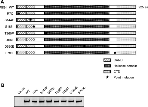
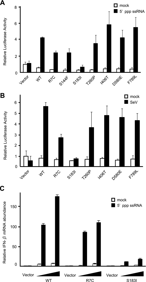
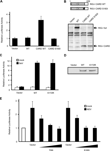
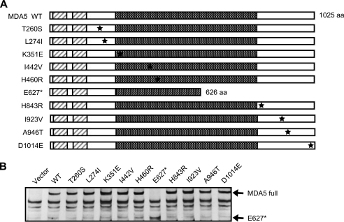
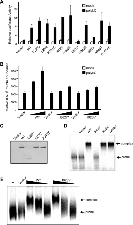
Retinoic acid-inducible gene I (RIG-I) and melanoma differentiation-associated gene 5 (MDA5) are essential for detecting viral RNA and triggering antiviral responses, including production of type I interferon. We analyzed the phenotype of non-synonymous mutants of human RIG-I and MDA5 reported in databases by functional complementation in cell cultures. Of seven missense mutations of RIG-I, S183I, which occurs within the second caspase recruitment domain repeat, inactivated this domain and conferred a dominant inhibitory function. Of 10 mutants of MDA5, two exhibited loss of function. A nonsense mutation, E627*, resulted in deletion of the C-terminal region and double-stranded RNA (dsRNA) binding activity. Another loss of function mutation, I923V, which occurs within the C-terminal domain, did not affect dsRNA binding activity, suggesting a novel and essential role for this residue in the signaling. Remarkably, these mutations are implicated in resistance to type I diabetes. However, the A946T mutation of MDA5, which has been implicated in type I diabetes by previous genetic analyses, affected neither dsRNA binding nor IFN gene activation. These results provide new insights into the structure-function relationship of RIG-I-like receptors as well as into human RIG-I-like receptor polymorphisms, antiviral innate immunity, and autoimmune diseases.


Figures

FIGURE 1.
FIGURE 1.
RIG-I nsSNP mutants and their expression in MEFs. A, schematic representation of the RIG-I wild type and nsSNP-containing mutants. RIG-I has a tandem CARD, RNA helicase domain, and CTD. Positions of the mutations are indicated by asterisks. aa, amino acids. B, FLAG-tagged WT RIG-I and SNPs were produced in RIG-I–/– MEFs and detected by immunoblotting using an anti-FLAG antibody.
FIGURE 2.
FIGURE 2.
Functional analysis of RIG-I nsSNP mutants. A and B, RIG-I–/– MEF cells were transiently transfected with p-55CIBluc together with empty vector (Vector) or the indicated constructs. The cells were subjected to a Dual-Luciferase assay after stimulation with 5′-ppp-ssRNA (12 h) (A) or SeV (12 h) (B). The relative firefly luciferase activity, normalized to the Renilla luciferase activity, is shown. Error bars show the SDs for triplicate transfections. mock, mock-treated. C, RIG-I–/– MEFs were transfected with empty vector (Vector) or expression vectors for WT RIG-I or mutants as indicated (the total amount of plasmid was kept at 6μgby adding empty vector). To observe the dose response, cells were transfected with 3 or 6 μg of the expression plasmid. Cells were mock-treated or transfected with 5′-ppp-ssRNA for 12 h, and IFN-β mRNA was quantified by quantitative PCR by using the Applied Biosystems primer set for mouse interferon-β1: Mm00439546_S1.
FIGURE 3.
FIGURE 3.
Characterization of RIG-I S183I. A, RIG-I–/– MEF cells were transfected with reporter genes together with empty vector (Vector) or plasmid expressing FLAG-tagged WT RIG-I, RIG-I CARD (the N-terminal region, amino acid 1–229), or RIG-I CARD S183I. After transfection (24 h), the cells were subjected to a Dual-Luciferase assay. Error bars show the S.D. values for triplicate transfections. B, each protein expressed in RIG-I–/– MEF cells was detected by immunoblotting using an anti-FLAG antibody. RIG-I full, full-length RIG-I. C, reporter assay of the K172R mutant was performed as in Fig. 2B. D, protein levels were determined by immunoblotting. RIG-I K172R and WT RIG-I were expressed at comparable levels. E, empty vector or expression vectors for full-length RIG-I with the T55I or S183I mutation were introduced into L929 cells (the total amount of plasmid was kept at 9 μg by adding empty vector) and infected with Newcastle disease virus, and reporter activity was analyzed as in panel A. To observe the dose response, cells received 1, 5, or 9 μg of the expression plasmid for T55I and S183I as indicated. NDV, Newcastle disease virus.
FIGURE 4.
FIGURE 4.
MDA5 nsSNP mutants and their expression in MEFs. A, schematic representation of WT MDA5 and its nsSNPs. Point mutations are indicated by asterisks. E627* is the nonsense mutant. aa, amino acids. B, FLAG-tagged MDA5 SNPs were produced in MDA5–/– MEFs and detected by immunoblotting using an anti-FLAG antibody. Vector, empty vector; MDA5 full, full-length MDA5.
FIGURE 5.
FIGURE 5.
Functional analysis of MDA5 nsSNP mutants. A, MDA5–/– MEFs were transfected with reporter genes together with the indicated constructs as in Fig. 2A. After stimulation with poly(I-C) (12 h), the cells were subjected to a Dual-Luciferase assay. Error bars show the S.D. values for triplicate transfections. mock, mock-treated. Vector, empty vector. B, MDA5–/– MEFs were transfected with expression vectors for WT MDA5 or E627* or Ile-923 mutants (the total amount of plasmid was kept at 6 μg by adding empty vector). To observe the dose response, cells were transfected with 3 or 5.7 μg of the expression plasmid. Cells were mock-treated or transfected with poly(I-C) for 12 h, and IFN-β mRNA was quantified by quantitative PCR as in Fig. 2C. C, 293T cells were transfected with empty vector, WT MDA5, E627*, I923V, or A946T, and the produced proteins were purified using anti-FLAG (“Experimental Procedures”). The purified proteins were separated by SDS-PAGE and stained by Coomassie Brilliant Blue. D, EMSA of the purified MDA5 proteins (500 ng) using 32P-labeled poly(I-C) as a probe. complex, probe protein complex; probe, free probe. E, dose response of RNA binding by WT MDA5 and the I923V mutant. EMSA was performed using 500, 300, and 100 ng of MDA5 and Ile-923 protein.

References

    1. Samuel, C. E. (2001) Clin. Microbiol. Rev. 14 778–809 - PMC - PubMed
    1. Akira, S., Uematsu, S., and Takeuchi, O. (2006) Cell 124 783–801 - PubMed
    1. Yoneyama, M., Kikuchi, M., Natsukawa, T., Shinobu, N., Imaizumi, T., Miyagishi, M., Taira, K., Akira, S., and Fujita, T. (2004) Nat. Immun. 5 730–737 - PubMed
    1. Yoneyama, M., and Fujita, T. (2008) Immunity 29 178–181 - PubMed
    1. Cui, S., Eisenacher, K., Kirchhofer, A., Brzozka, K., Lammens, A., Lammens, K., Fujita, T., Conzelmann, K. K., Krug, A., and Hopfner, K. P. (2008) Mol. Cell 29 169–179 - PubMed

Publication types