. 2009 Jun;58(6):1391-402.
doi: 10.2337/db08-1697. Epub 2009 Mar 26.

Inactivation of GSK-3beta by metallothionein prevents diabetes-related changes in cardiac energy metabolism, inflammation, nitrosative damage, and remodeling

Affiliations

Inactivation of GSK-3beta by metallothionein prevents diabetes-related changes in cardiac energy metabolism, inflammation, nitrosative damage, and remodeling

Yuehui Wang et al. Diabetes. 2009 Jun.

Abstract

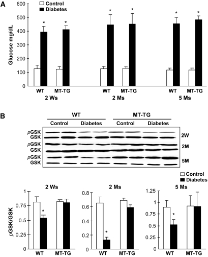
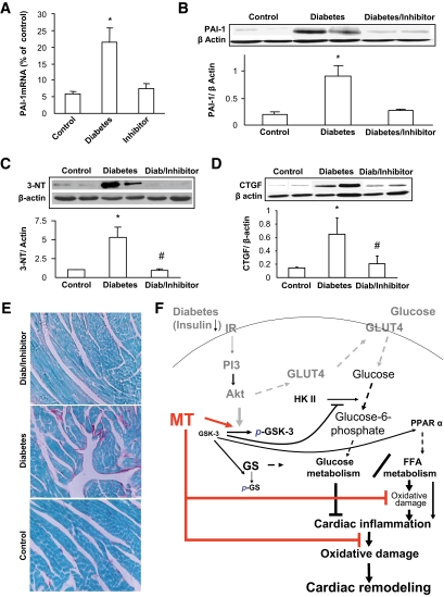
Objective: Glycogen synthase kinase (GSK)-3beta plays an important role in cardiomyopathies. Cardiac-specific metallothionein-overexpressing transgenic (MT-TG) mice were highly resistant to diabetes-induced cardiomyopathy. Therefore, we investigated whether metallothionein cardiac protection against diabetes is mediated by inactivation of GSK-3beta.

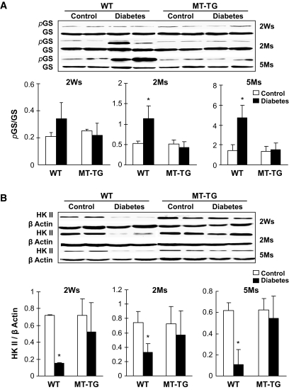
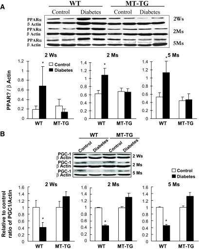
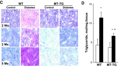
Research design and methods: Diabetes was induced with streptozotocin in both MT-TG and wild-type mice. Changes of energy metabolism-related molecules, lipid accumulation, inflammation, nitrosative damage, and fibrotic remodeling were examined in the hearts of diabetic mice 2 weeks, 2 months, and 5 months after the onset of diabetes with Western blotting, RT-PCR, and immunohistochemical assays.

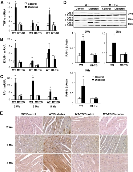
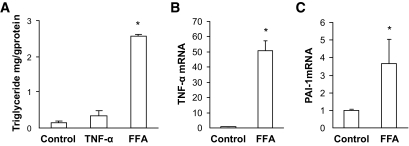
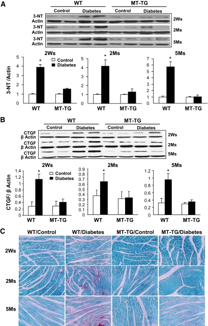
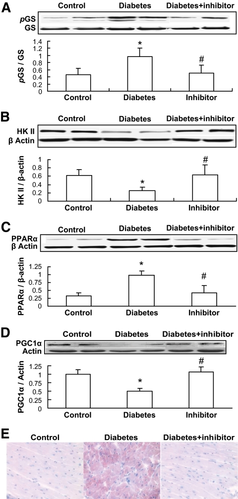
Results: Activation (dephosphorylation) of GSK-3beta was evidenced in the hearts of wild-type diabetic mice but not MT-TG diabetic mice. Correspondingly, cardiac glycogen synthase phosphorylation, hexokinase II, PPARalpha, and PGC-1alpha expression, which mediate glucose and lipid metabolisms, were significantly changed along with cardiac lipid accumulation, inflammation (TNF-alpha, plasminogen activator inhibitor 1 [PAI-1], and intracellular adhesion molecule 1 [ICAM-1]), nitrosative damage (3-nitrotyrosin accumulation), and fibrosis in the wild-type diabetic mice. The above pathological changes were completely prevented either by cardiac metallothionein in the MT-TG diabetic mice or by inhibition of GSK-3beta activity in the wild-type diabetic mice with a GSK-3beta-specific inhibitor.

Conclusions: These results suggest that activation of GSK-3beta plays a critical role in diabetes-related changes in cardiac energy metabolism, inflammation, nitrosative damage, and remodeling. Metallothionein inactivation of GSK-3beta plays a critical role in preventing diabetic cardiomyopathy.


Figures

FIG. 1.
FIG. 1.
Diabetic hyperglycemia and activation of GSK-3β. Before mice were killed at the indicated times, blood glucose was monitored at indicated times, using tail-vein blood (A), and cardiac tissues were used for Western blotting analysis of total and phosphorylated GSK-3β (B). *P < 0.05 vs. corresponding controls. Ms, months; Ws, weeks; WT, wild type.
FIG. 2.
FIG. 2.
Diabetes-induced glycogen synthase (GS) phosphorylation and decrease of HK II expression. Cardiac tissues were collected, as indicated in Fig. 1, for detecting total and phosphorylated glycogen synthase (Ser641) (A) and HK II expression (B) by Western blotting. *P < 0.05 vs. control. Ms, months; Ws, weeks; WT, wild type.
FIG. 3.
FIG. 3.
Diabetes-increased PPARα expression and lipid accumulation. Cardiac tissues were collected, as indicated in Fig. 1, for detecting PPARα (A) and PGC-1α (B) expressions by Western blotting and cardiac lipid accumulation by Oil Red O staining (400×) (C) and triglyceride measurement (D). Panel D presents the data only from diabetic mice 2 weeks (Ws) after diabetes. *P < 0.05 vs. control; #P < 0.05 vs. wild-type (WT) diabetic group. Ms, months. (A high-quality digital representation of this figure is available in the online issue.).
FIG. 3.
FIG. 3.
Diabetes-increased PPARα expression and lipid accumulation. Cardiac tissues were collected, as indicated in Fig. 1, for detecting PPARα (A) and PGC-1α (B) expressions by Western blotting and cardiac lipid accumulation by Oil Red O staining (400×) (C) and triglyceride measurement (D). Panel D presents the data only from diabetic mice 2 weeks (Ws) after diabetes. *P < 0.05 vs. control; #P < 0.05 vs. wild-type (WT) diabetic group. Ms, months. (A high-quality digital representation of this figure is available in the online issue.).
FIG. 4.
FIG. 4.
Diabetes-induced cardiac inflammation. Real-time RT-PCR was used to examine the expression of TNF-α (A), ICAM-1 (B), and PAI-1 mRNA (C) in the hearts of diabetic mice at indicated postdiabetic times. TNF-α, ICAM-1, and PAI-1 expressions were normalized to GAPDH, by which the fold changes in expression were calculated using 2−ΔΔCt method. Expression of PAI-1 was further confirmed for its protein level by Western blotting (D) and localization by immunohistochemical staining (E). *P < 0.05 vs. corresponding controls. Ms, months; Ws, weeks; WT, wild type. (A high-quality digital representation of this figure is available in the online issue.)
FIG. 5.
FIG. 5.
FFA-induced upregulation of inflammatory cytokines. Cultured cardiac (H9c2) cells were directly exposed to TNF-α at a concentration of 1.0 ng/ml for 24 h, which did not increase intracellular triglyceride levels (A). Direct exposure of cardiac cells to FFA (palmitate) at 50 μmol/l for 24 h significantly increased intracellular triglyceride levels (A) and also significantly increased mRNA expression of inflammatory cytokines TNF-α (B) and PAI-1 (C). *P < 0.05 vs. control.
FIG. 6.
FIG. 6.
Diabetes-increased cardiac nitrosative damage and fibrosis. Cardiac nitrosative damage was examined by 3-NT accumulation with Western blotting analysis (A). Cardiac fibrosis was examined by Western blotting analysis of CTGF expression (B) and Sirius Red staining of collagen (C). *P < 0.05 vs. corresponding controls. Ms, months; Ws, weeks; WT, wild type. (A high-quality digital representation of this figure is available in the online issue.)
FIG. 7.
FIG. 7.
Inhibition of GSK-3β by its inhibitor attenuated diabetes-induced changes related to glucose and lipid metabolism. Once diabetes was diagnosed on day 3 after mice were injected with STZ, half of these diabetic mice were immediately administered GSK-3β–specific inhibitor SB216763 at 600 μg/kg every other day for 2 months, and then cardiac glycogen synthase (GS) phosphorylation (A), HK II expression (B), PPARα expression (C), and PGC-1α expression (D) were examined by Western blotting. Cardiac lipid accumulation was examined by Oil Red O staining (400×) (E). *P < 0.05 vs. corresponding controls; #P < 0.05 vs. corresponding diabetes. (A high-quality digital representation of this figure is available in the online issue.)
FIG. 8.
FIG. 8.
Inhibition of GSK by its inhibitor attenuated diabetes-induced cardiac inflammation, nitrosative damage, and fibrosis. Experimental approaches and cardiac tissue sampling are the same as those in Fig. 6. Samples were examined for cardiac expression of PAI-1 by real-time RT-PCR (A) and Western blotting (B), cardiac 3-NT accumulation (C), and cardiac fibrosis by Western blotting for CTGF expression (D) and Sirius Red staining of collagen (200×) (E). *P < 0.05 vs. corresponding controls; #P < 0.05 vs. corresponding diabetes. F: schematic illustration of the mechanisms by which metallothionein (MT) preserves GSK-3β phosphorylation to maintain glucose and lipid metabolism balance under diabetic conditions and, consequently, inhibit cardiac lipid accumulation, inflammation, oxidative/nitrosative damage, and remodeling. Solid lines present the experimental finding from the present study, and dashed lines indicate well-known pathways from the literature. GS, glycogen synthase; IR, insulin receptor. (A high-quality digital representation of this figure is available in the online issue.)

References

    1. Ye G, Metreveli NS, Ren J, Epstein PN: Metallothionein prevents diabetes-induced deficits in cardiomyocytes by inhibiting reactive oxygen species production. Diabetes 2003; 52: 777– 783 - PubMed
    1. Cai L, Wang J, Li Y, Sun X, Wang L, Zhou Z, Kang YJ: Inhibition of superoxide generation and associated nitrosative damage is involved in metallothionein prevention of diabetic cardiomyopathy. Diabetes 2005; 54: 1829– 1837 - PubMed
    1. Fang CX, Dong F, Ren BH, Epstein PN, Ren J: Metallothionein alleviates cardiac contractile dysfunction induced by insulin resistance: role of Akt phosphorylation, PTB1B, PPARgamma and c-Jun. Diabetologia 2005; 48: 2412– 2421 - PubMed
    1. Cai L, Wang Y, Zhou G, Chen T, Song Y, Li X, Kang YJ: Attenuation by metallothionein of early cardiac cell death via suppression of mitochondrial oxidative stress results in a prevention of diabetic cardiomyopathy. J Am Coll Cardiol 2006; 48: 1688– 1697 - PubMed
    1. Dong F, Li Q, Sreejayan N, Nunn JM, Ren J: Metallothionein prevents high-fat diet–induced cardiac contractile dysfunction: role of peroxisome proliferator–activated receptor γ coactivator 1α and mitochondrial biogenesis. Diabetes 2007; 56: 2201– 2212 - PubMed

Publication types

MeSH terms