. 2009 May 8;324(5928):797-801.
doi: 10.1126/science.1172482. Epub 2009 Mar 26.

Human induced pluripotent stem cells free of vector and transgene sequences

Affiliations

Human induced pluripotent stem cells free of vector and transgene sequences

Junying Yu et al. Science. .

Abstract

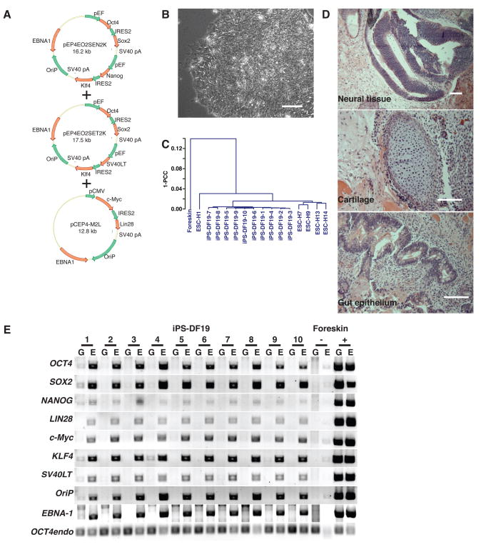
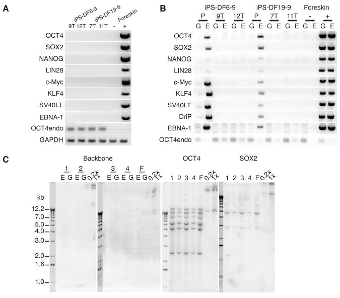
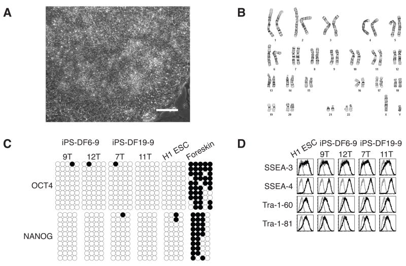
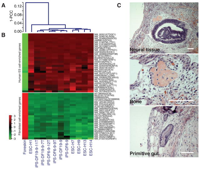
Reprogramming differentiated human cells to induced pluripotent stem (iPS) cells has applications in basic biology, drug development, and transplantation. Human iPS cell derivation previously required vectors that integrate into the genome, which can create mutations and limit the utility of the cells in both research and clinical applications. We describe the derivation of human iPS cells with the use of nonintegrating episomal vectors. After removal of the episome, iPS cells completely free of vector and transgene sequences are derived that are similar to human embryonic stem (ES) cells in proliferative and developmental potential. These results demonstrate that reprogramming human somatic cells does not require genomic integration or the continued presence of exogenous reprogramming factors and removes one obstacle to the clinical application of human iPS cells.


Figures

Fig. 1
Fig. 1
Reprogramming human foreskin fibroblasts without genomic vector integration. (A) Episomal vectors (combination 19 from experiment 3, Table S2). pEF: the eukaryotic elongation factor 1α promoter; pCMV: the cytomegalovirus immediate-early promoter. Transgenes and other features of vectors are indicated by red and green arrows respectively. (B) Bright-field image of iPS cells obtained by transfection of combination 19 episomal vectors (clone DF19-9: “Defined Factor” combination 19, and clone 9). Scale bars, 0.1 mm. (C) Pearson correlation analyses of global gene expression (51,337 transcripts) in human fibroblast-derived iPS cell clones (combination 19). 1-PCC: Pearson Correlation Coefficient. (D) Hematoxylin and eosin staining of teratoma sections of iPS cell clone DF19-9 (53 days after injection). Teratomas were obtained from all ten iPS-DF19 clones. Scale bars, 0.1 mm. (E) PCR analysis of episomal DNA in iPS-DF19 clone 1 to 10. G: genomic DNA template; E: episomal DNA template. Genomic and episomal DNA from nontransfected and combination 19 episomal vector-transfected (day 17 posttransfection) fibroblasts were used as negative (−) and positive (+) controls respectively. 32 PCR cycles were used for all primer sets.
Fig. 2
Fig. 2
Human foreskin fibroblast-derived iPS cells free of vectors and transgenes. (A) RT-PCR analysis of transgene expression in iPS-DF6-9 subclone 9T and 12T, and iPS-DF-19-9 subclone 7T and 11T. Negative control (−): fibroblasts. Positive control (+): fibroblasts transfected with combination 19 episomal vectors (day 4 posttransfection). 32 PCR cycles were used for all primer sets. (B) PCR analysis of episomal DNA in iPS-DF6-9 (P: parental clone), iPS-DF6-9 subclone 9T and 12T, iPS-DF19-9 (P), and iPS-DF19-9 subclones 7T and 11T. G: genomic DNA template; E: episomal DNA template. Negative (−) and positive (+) controls were the same as in Fig. 1E. 32 PCR cycles were used for all primer sets except OCT4endo (28 cycles). (C) Southern blot analysis of exogenous DNA in iPS-DF6-9 and iPS-DF-19-9 subclones. The pCEP4 vector was used as a probe to detect the presence of vector backbone, and the open reading frames of OCT4 and SOX2 were used as probes to examine both the endogenous gene and possible transgenes. 1: iPS-DF6-9-9T; 2: iPS-DF6-9-12T; 3: iPS-DF19-9-7T; 4: iPS-DF19-9-11T; F: foreskin fibroblasts. E: undigested episomal DNA; G: digested genomic DNA. Combination 19 episomal vector DNA diluted to the equivalents of 0.2 and 1 integration per genome was used as positive controls (0.2x and 1x).
Fig. 3
Fig. 3
Characterization of iPS cell subclones. (A) Bright-field image of iPS-DF6-9-12T. Scale bar, 0.1 mm. (B) G-banding chromosome analysis of iPS-DF6-9-12T. (C) Analysis of the methylation status of the OCT4 and NANOG promoters in iPS cell subclones using bisulfite sequencing. Open circles indicate unmethylated, and filled circles indicate methylated CpG dinucleotides. (D) Flow cytometry expression analysis of human ES cell-specific cell surface markers. Gray line: isotype control; black line: antigen staining.
Fig. 4
Fig. 4
Characterization of iPS cell subclones. (A) Pearson correlation analyses of global gene expression (51,337 transcripts) in iPS cell parental clone DF6-9 and DF19-9; iPS cell subclone DF6-9-9T, DF6-9-12T, DF19-9-7T, and DF19-9-11T; five human ES cell lines; foreskin fibroblasts. 1-PCC: Pearson Correlation Coefficient. (B) Expression of genes that are differentially expressed between human ES cells and foreskin fibroblasts. Top panel: 30 well-known human ES cell-enriched genes; bottom panel: top 25 foreskin fibroblast-enriched genes. The color key is shown on the left. (C) Hematoxylin and eosin staining of teratoma sections of iPS-DF19-9-11T (7 weeks after injection). Teratomas were obtained from all four iPS cell subclones. Scale bars, 0.1 mm.

References

    1. Thomson JA, et al. Science. 1998;282:1145. - PubMed
    1. Yu J, et al. Science. 2007;318:1917. - PubMed
    1. Takahashi K, et al. Cell. 2007;131:861. - PubMed
    1. Takahashi K, Yamanaka S. Cell. 2006;126:663. - PubMed
    1. Okita K, Ichisaka T, Yamanaka S. Nature. 2007;448:313. - PubMed

Publication types

Substances

Associated data