. 2009 Aug;30(8):1281-7.
doi: 10.1093/carcin/bgp071. Epub 2009 Mar 26.

The type III transforming growth factor-beta receptor negatively regulates nuclear factor kappa B signaling through its interaction with beta-arrestin2

Affiliations

The type III transforming growth factor-beta receptor negatively regulates nuclear factor kappa B signaling through its interaction with beta-arrestin2

Hye Jin You et al. Carcinogenesis. 2009 Aug.

Abstract

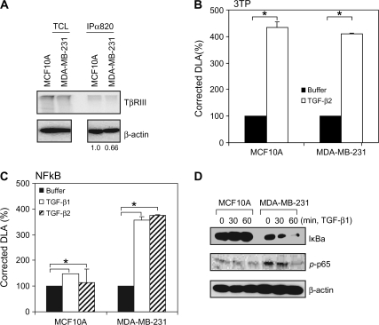
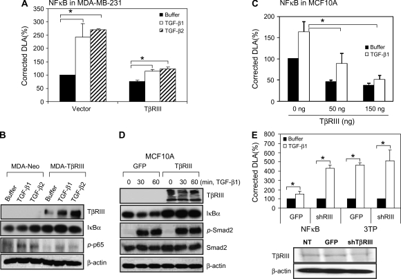
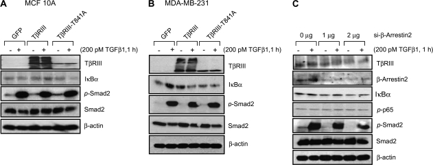
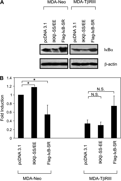
Transforming growth factor-beta (TGF-beta) increases or decreases nuclear factor kappa B (NFkappaB) signaling in a context-dependent manner through mechanisms that remain to be defined. The type III transforming growth factor-beta receptor (TbetaRIII) is a TGF-beta superfamily co-receptor with emerging roles in both mediating and regulating TGF-beta superfamily signaling. We have previously reported a novel interaction of TbetaRIII with the scaffolding protein, beta-arrestin2, which results in TbetaRIII internalization and downregulation of TGF-beta signaling. beta-arrestin2 also scaffolds interacting receptors with the mitogen-activated protein kinase and NFkappaB-signaling pathways. Here, we demonstrate that TbetaRIII, through its interaction with beta-arrestin2, negatively regulates NFkappaB signaling in MCF10A breast epithelial and MDA-MB-231 breast cancer cells. Increasing TbetaRIII expression reduced NFkappaB-mediated transcriptional activation and IkappaBalpha degradation, whereas a TbetaRIII mutant unable to interact with beta-arrestin2, TbetaRIII-T841A, had no effect. In a reciprocal manner, short hairpin RNA-mediated silencing of either TbetaRIII expression or beta-arrestin2 expression increased NFkappaB-mediated transcriptional activation and IkappaBalpha degradation. Functionally, TbetaRIII-mediated repression of NFkappaB signaling is important for TbetaRIII-mediated inhibition of breast cancer cell migration. These studies define a mechanism through which TbetaRIII regulates NFkappaB signaling and expand the roles of this TGF-beta superfamily co-receptor in regulating epithelial cell homeostasis.


Figures

Fig. 1.
Fig. 1.
TβRIII expression correlates with NFκB signaling in MCF10A and MDA-MB-231 cells. (A) Cell surface expression of TβRIII in MCF10A and MDA-MB-231 cells was demonstrated by binding and cross-linking assay as described in Materials and Methods. Both total cell lysate and immunoprecipitates with anti-TβRIII antibody (820) were analyzed. For the immunoprecipitates, densitometry of TβRIII expression controlled for β-actin is presented below. (B and C) Cells were transfected with pNFκB-Luc (B) or p3TP-Luc (C) and pRL-SV40 using Fugene 6 and grown for 24 h. Cells were treated with buffer, TGF-β1 (100 pM) or TGF-β2 (200 pM) for 24 h and harvested for luciferase activity assay. Data are expressed as means ± SDs of at least three independent experiments. Statistical significance of differences was assessed using unpaired Student's t-tests (*P < 0.01). (D) Subconfluent cells were treated with TGF-β1 (200 pM) for the indicated times and harvested for western blotting with the indicated antibodies. The results shown are representative of at least three independent experiments.
Fig. 2.
Fig. 2.
TβRIII negatively regulates NFκB signaling. (A and C) MCF10A and MDA-MB-231 cells were transfected with pNFκB-Luc, pRL-SV40 and TβRIII (300 ng per well in a 24-well plate for MDA-MB-231 cells, indicated amounts for MCF10A cells) using Fugene 6 and grown for 24 h. Cells were treated with buffer, TGF-β1 (100 pM) or TGF-β2 (200 pM) for 24 h and harvested for luciferase activity assay. Data are expressed as means ± SDs of at least three independent experiments. Statistical significance of differences was assessed using unpaired Student's t-tests (*P < 0.01). (B) MDA-MB-231-Neo and MDA-MB-231-TβRIII cells were treated with TGF-β1 or TGF-β2 for 1 h, harvested and subjected to western blotting. (D) Subconfluent MCF10A cells were infected with Adeno-TβRIII or GFP-adenovirus. After 2 days, cells were treated with TGF-β1 (200 pM) for the indicated times and harvested for western blotting with the indicated antibodies. The results shown are representative of at least three independent experiments. (E) MCF10A cells were infected with GFP or shRNA of TβRIII adenovirus and transfected with pNFκB and pRL-SV40 for reporter gene assay. After 1 day, cells were treated with TGF-β1 or TGF-β2 for 24 h and subjected to reporter gene assay and binding and cross-linking assay. Data are expressed as means ± SDs of at least three independent experiments. Statistical significance of differences was assessed using unpaired Student's t-tests (*P < 0.01).
Fig. 3.
Fig. 3.
TβRIII negatively regulates NFκB signaling via β-arrestin2. MCF10A (A) and MDA-MB-231 (B) cells were infected with adenovirus expressing GFP, TβRIII or TβRIII-T841A and grown for 48 h. Cells were then treated with TGF-β1, 200 pM for 1 h, harvested and subjected to western blotting with the indicated antibodies. (C) MDA-MB-231 cells were transfected with siRNA against β-arrestin2 for 2 days and treated with TGF-β1 (200 pM, 1 h), after which cells were harvested for western blotting. The results shown are representative of at least three independent experiments.
Fig. 4.
Fig. 4.
TβRIII suppresses cell migration, in part, by negatively regulating the NFκB pathway. MDA-MB-231 cells were transfected with pcDNA3.1, IKKβ-SS/EE or IκB-super-repressor (SR) and grown for 48 h. Cells were then harvested for western blotting (A) or migration assays (B). The results shown are the average of four independent experiments ± SEM, NS, not significant, *P< 0.05.

References

    1. Bierie B, et al. Tumour microenvironment: TGFbeta: the molecular Jekyll and Hyde of cancer. Nat. Rev. Cancer. 2006;6:506–520. - PubMed
    1. Elliott RL, et al. Role of transforming growth factor Beta in human cancer. J. Clin. Oncol. 2005;23:2078–2093. - PubMed
    1. Gordon KJ, et al. Role of transforming growth factor-beta superfamily signaling pathways in human disease. Biochim. Biophys. Acta. 2008;1782:197–228. - PubMed
    1. Kirkbride KC, et al. Cell-surface co-receptors: emerging roles in signaling and human disease. Trends Biochem. Sci. 2005;30:611–621. - PubMed
    1. Massague J, et al. TGFbeta signaling in growth control, cancer, and heritable disorders. Cell. 2000;103:295–309. - PubMed

Publication types

MeSH terms