. 2009;4(3):e4839.
doi: 10.1371/journal.pone.0004839. Epub 2009 Mar 27.

Expression analysis of the Theileria parva subtelomere-encoded variable secreted protein gene family

Affiliations

Expression analysis of the Theileria parva subtelomere-encoded variable secreted protein gene family

Jacqueline Schmuckli-Maurer et al. PLoS One. 2009.

Abstract

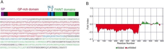
Background: The intracellular protozoan parasite Theileria parva transforms bovine lymphocytes inducing uncontrolled proliferation. Proteins released from the parasite are assumed to contribute to phenotypic changes of the host cell and parasite persistence. With 85 members, genes encoding subtelomeric variable secreted proteins (SVSPs) form the largest gene family in T. parva. The majority of SVSPs contain predicted signal peptides, suggesting secretion into the host cell cytoplasm.

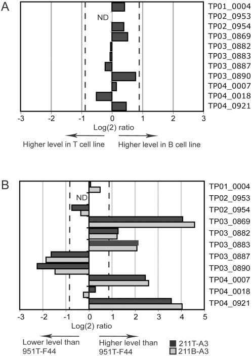
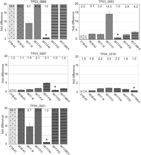
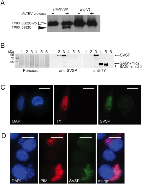
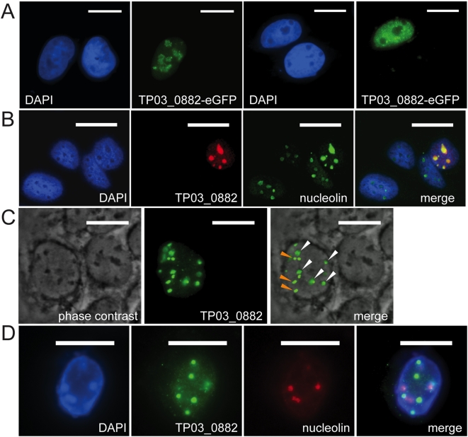
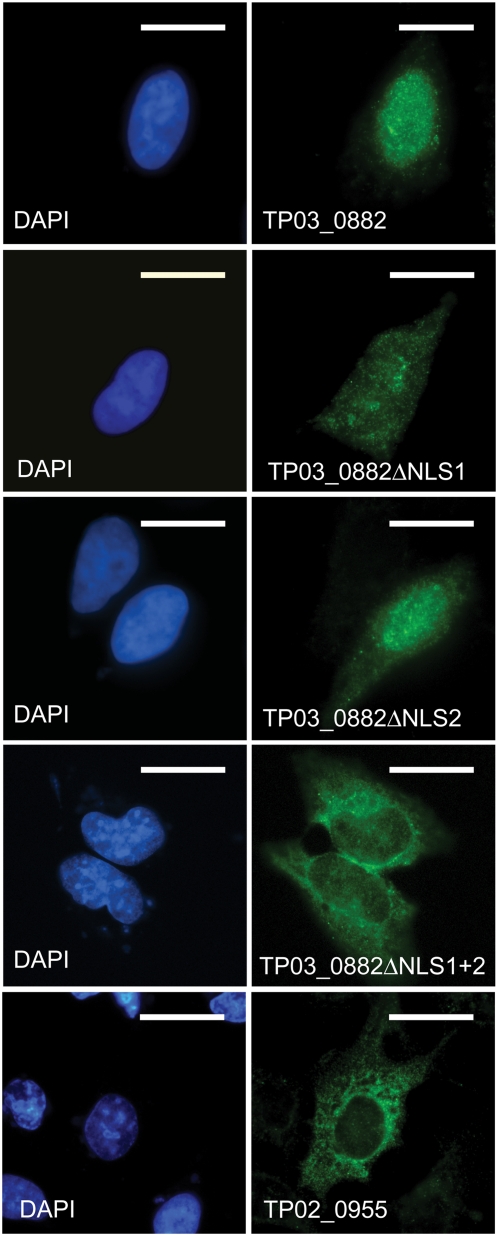
Methodology/principal findings: We analysed SVSP expression in T. parva-transformed cell lines established in vitro by infection of T or B lymphocytes with cloned T. parva parasites. Microarray and quantitative real-time PCR analysis revealed mRNA expression for a wide range of SVSP genes. The pattern of mRNA expression was largely defined by the parasite genotype and not by host background or cell type, and found to be relatively stable in vitro over a period of two months. Interestingly, immunofluorescence analysis carried out on cell lines established from a cloned parasite showed that expression of a single SVSP encoded by TP03_0882 is limited to only a small percentage of parasites. Epitope-tagged TP03_0882 expressed in mammalian cells was found to translocate into the nucleus, a process that could be attributed to two different nuclear localisation signals.

Conclusions: Our analysis reveals a complex pattern of Theileria SVSP mRNA expression, which depends on the parasite genotype. Whereas in cell lines established from a cloned parasite transcripts can be found corresponding to a wide range of SVSP genes, only a minority of parasites appear to express a particular SVSP protein. The fact that a number of SVSPs contain functional nuclear localisation signals suggests that proteins released from the parasite could contribute to phenotypic changes of the host cell. This initial characterisation will facilitate future studies on the regulation of SVSP gene expression and the potential biological role of these enigmatic proteins.


Conflict of interest statement

Competing Interests: The authors have declared that no competing interests exist.

Figures

Figure 1
Figure 1. Schematic presentation of TP08_0882, a typical SVSP.
A. Schematic view and amino acid sequence of TP03_0882. TP03_0882 is 607 amino acids long. The polypeptide has a putative signal peptide (SP) for secretion (purple) with a cleavage site after residue 21 (predicted by the SignalP3.0 web server). A large N-terminal region containing abundant Q and P residues (red) is followed by C-terminal region containing two nuclear localisation signals (NLS 1 and 2, blue) and two FAINT domains from aa 422 to 481 and aa 520 to 579 (green). B. Analysis of the TP03_0882 protein using the FoldIndex© software. The N-terminal region of the protein is intrinsically unfolded, while the conserve C-terminal region is predicted to fold.
Figure 2
Figure 2. Quantitative RT-PCR analysis of SVSP expression patterns in different T. parva-infected cell lines.
A. Comparison of SVSP transcript levels in T and B cell lines transformed with the T. parva (Marikebuni) A3 clone. Cells originated from the same animal (211). Relative transcript levels for 11 selected SVSP genes (listed on the right) were assessed by qRT-PCR in the cell lines 211T-A3 and 211B-A3. Dashed lines indicate log(2) ratios of 0.9 and -0.9, the arbitrarily defined Index threshold used for microarray analysis above which differential expression of genes is considered significant. For one gene no transcripts were detected (ND). B. SVSP transcript levels in the T. parva (Marikebuni) A3-infected cell lines 211T-A3 (dark bars) and 211B-A3 (pale bars) compared to a third cell line 951T-F44, infected with a different T. parva (Marikebuni) clone (F44).
Figure 3
Figure 3. qRT-PCR analysis of five SVSP genes in seven different cloned T. parva–infected cell lines.
The expression of five SVSP genes, TP03_0869, TP03_0883, TP03_0887, TP04_0018 and TP04_0921 was monitored in seven cell lines transformed by different T. parva clones (see Table 2 for cell lines). For each gene and each cell line, the relative fold difference in relation to the cell line with the lowest transcript level of the respective gene (indicated as 1.0, labeled with *) is indicated. Data represent the mean of the transcript levels from two independent culture flasks. The actual fold difference is indicated at the top of the bar. Error bars denote the standard error of the mean. Differently coloured bars indicate cell lines from different animals. Dotted bars indicate B cell lines, bars with no pattern indicate γδ T cells, and CD4+ T cells are indicated with striped bars.
Figure 4
Figure 4. Detection of recombinant and parasite SVSP by immunoblot and immunofluorescence analysis.
A. The C-terminal region of SVSP TP03_0882 was expressed as a V5/His-epitope tagged protein in bacteria and subjected to immunoblot analysis using antibodies raised in rats against the same polypeptide (anti-SVSP) or anti-V5 antibodies. Open arrowhead indicates the uncleaved protein; closed arrowhead indicates C-terminal TP03_0882 after removal of the V5/His epitope tag by AcTEV protease. B. BoMac cells were transfected with untagged and TY-tagged full length TP03_0882 and analysed by immunoblot, using anti-SVSP or anti-TY antibodies. The position of SVSP and two TY-tagged control proteins SAG1-mic2 and SAG1-mic20 are indicated. A Ponceau-stained filter is added for loading control and also shows the position of the molecular weight markers. line 1: BoMac transfected with pmaxGFP (Amaxa); line2: BoMac transfected with pmaxCloning-TP03_0882; line 3: BoMac transfected with pmax-TP03_0882TY; line 4: BoMac transfected with pcDNA-SAG1-Ty-MIC2; line 5: BoMac transfected with pCAN-SAG1-Ty-MIC20; line 6: Whole cell lysate of T. parva (Muguga)- infected cells (TpMD409 CD8+ T cell). C. Detection of TP03_0882 SVSP expressed as a TY-tagged protein in BoMac cells. The nuclei of two cells are stained by DAPI. The cell on the left expresses SVSP as indicated by its reactivity with TY antibodies. The cell is also labelled after staining with anti-SVSP antibodies, showing nuclear localisation of the expressed protein. Scale bar: 20 μm. D. Cytospin preparations of T. parva (Muguga)-transformed cells (TpMD409 CD8+ T cells) mixed with uninfected bovine control BL20 cells were fixed in paraformaldehyde and analyzed by indirect immunofluorescence using anti-SVSP antibodies (green) and anti-PIM antibodies (red). Host and parasite nuclei were stained with DAPI (blue). The control cell in the middle does not harbour a parasite and is negative for PIM and SVSP. Both the cells at the top and the bottom are parasitised and thus labelled with anti-PIM. Only the parasite in the cell at the bottom, reacts with antibodies raised against TP03_0882 SVSP (green) partly revealing a vesicular staining pattern. Merge is an overlay of the three panels. Scale bar: 10 μm
Figure 5
Figure 5. SVSP TP03_0882 expressed in U2OS cells localises to different nuclear compartments.
A. TP03_0882 was expressed as eGFP fusion protein in U2OS cells. Cells growing on coverslips were fixed with paraformaldehyde. DNA was stained with DAPI (blue). Of the two cells shown in the panels on the left, one expresses SVSP which is localised to the nucleoli. The panels on the right show a cell with a nucleoplasmic SVSP localisation pattern. Scale bar: 20 μM. B. Cells transfected with a plasmid encoding V5-tagged SVSP TP03_0882 were fixed with methanol and stained with antibodies directed against nucleolin (as nucleolar marker, green) or V5 (SVSP-expressing cell, red). The overlay (merge) reveals the colocalisation of SVSP and nucleolin in the nucleoli. Scale bar: 25 μm. C. In some transfected cells, SVSP (green) localises to nucleoli (white arrows) and other nuclear structures (orange arrows). In the left panel, nucleoli were visualized by phase contrast (dark areas). In the middle panel, SVSP is visualised using anti-V5 antibodies. Merge indicates the overlay of both panels. Scale bar: 20 μm. D. In some cells, SVSP (anti-V5, green) does not localise to the nucleoli (stained with anti-nucleolin, red) and is only found in dense nuclear bodies. Nuclei are stained blue (DAPI). Scale bar: 20 μm.
Figure 6
Figure 6. Identification of two functional NLS in SVSP TP03_0882.
U2OS cells were transfected with plasmid constructs encoding V5-tagged forms of TP03_0882 containing both NLS (TP03_0882), lacking NLS1 (TP03_0882ΔNLS1), lacking NLS2 (TP03_0882ΔNLS2) or both NLS (TP03_0882ΔNLS1+2). In addition, cells were also transfected with a plasmid encoding TP02_0995, an SVSP predicted not to contain a NLS. Cells were fixed in methanol and SVSP localisation monitored using anti-V5 antibodies (green). DNA was stained with DAPI (blue).

Similar articles

Cited by

References

    1. Barry JD, Ginger ML, Burton P, McCulloch R. Why are parasite contingency genes often associated with telomeres? International Journal for Parasitology. 2003;33:29–45. - PubMed
    1. Scherf A, Hernandez-Rivas R, Buffet P, Bottius E, Benatar C, et al. Antigenic variation in malaria: in situ switching, relaxed and mutually exclusive transcription of var genes during intra-erythrocytic development in Plasmodium falciparum. EMBO J. 1998;17:5418–5426. - PMC - PubMed
    1. Borst P, Ulbert S. Control of VSG gene expression sites. Mol Biochem Parasitol. 2001;114:17–27. - PubMed
    1. Filler SG. Candida-host cell receptor-ligand interactions. Curr Opin Microbiol. 2006;9:333–339. - PubMed
    1. Gottschling DE, Aparicio OM, Billington BL, Zakian VA. Position effect at S. cerevisiae telomeres: reversible repression of Pol II transcription. Cell. 1990;63:751–762. - PubMed

Publication types