. 2009;4(3):e5017.
doi: 10.1371/journal.pone.0005017. Epub 2009 Mar 27.

Cysteine redox potential determines pro-inflammatory IL-1beta levels

Affiliations

Cysteine redox potential determines pro-inflammatory IL-1beta levels

Smita S Iyer et al. PLoS One. 2009.

Abstract

Background: Cysteine (Cys) and its disulfide, cystine (CySS) represent the major extracellular thiol/disulfide redox control system. The redox potential (E(h)) of Cys/CySS is centered at approximately -80 mV in the plasma of healthy adults, and oxidation of E(h) Cys/CySS is implicated in inflammation associated with various diseases.

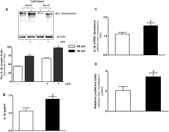
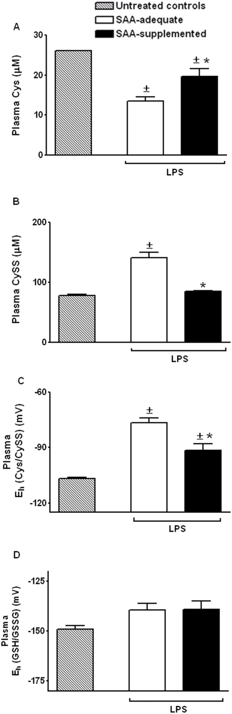
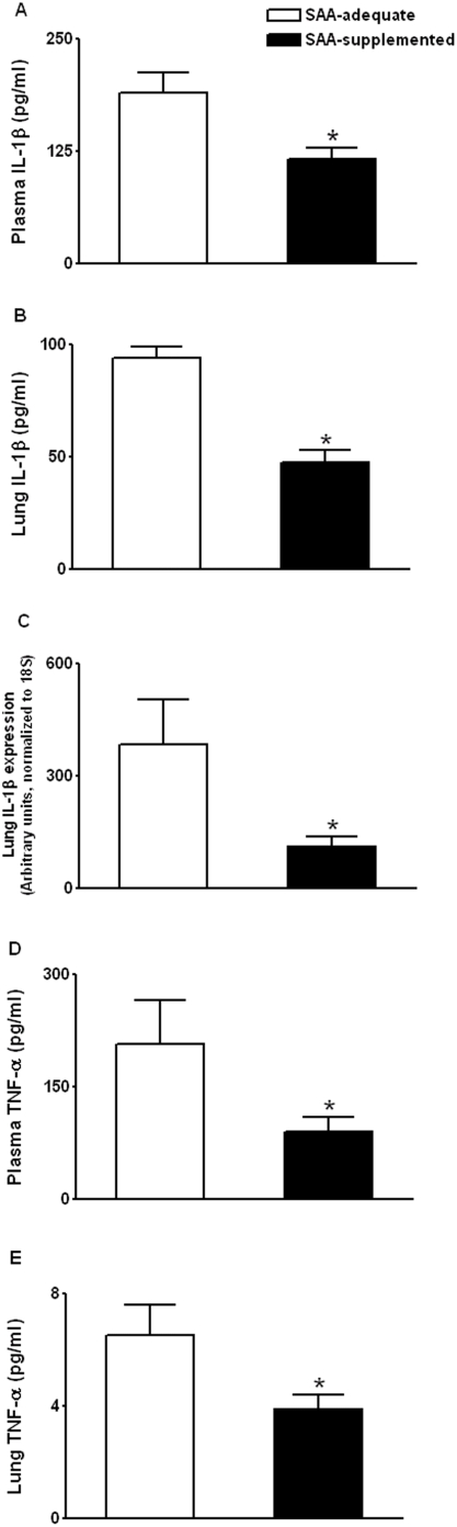
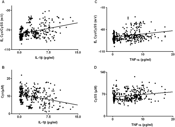
Methodology/principal findings: The purpose of the present study was to determine whether oxidized E(h) Cys/CySS is a determinant of interleukin (IL)-1beta levels. Results showed a 1.7-fold increase in secreted pro-IL-1beta levels in U937 monocytes exposed to oxidized E(h) Cys/CySS (-46 mV), compared to controls exposed to a physiological E(h) of -80 mV (P<0.01). In LPS-challenged mice, preservation of plasma E(h) Cys/CySS from oxidation by dietary sulfur amino acid (SAA) supplementation, was associated with a 1.6-fold decrease in plasma IL-1beta compared to control mice fed an isonitrogenous SAA-adequate diet (P<0.01). Analysis of E(h) Cys/CySS and IL-1beta in human plasma revealed a significant positive association between oxidized E(h) Cys/CySS and IL-1beta after controlling for age, gender, and BMI (P<0.001).

Conclusions/significance: These data show that oxidized extracellular E(h) Cys/CySS is a determinant of IL-1beta levels, and suggest that strategies to preserve E(h) Cys/CySS may represent a means to control IL-1beta in inflammatory disease states.


Conflict of interest statement

Competing Interests: The authors have declared that no competing interests exist.

Figures

Figure 1
Figure 1. Oxidized extracellular Eh Cys/CySS increases pro-IL-1β in monocytes.
U937 cells were exposed to physiological (−80 mV) and oxidized (−46 mV) Eh Cys/CySS for 8 h and levels of the IL-1β precursor were determined by Western blot (A). Western blot analysis of the cell extract revealed increase in the 31 kDa precursor form of IL-1β at −46 mV compared to –80 mV. Quantitative analysis of the band densities of three separate experiments is shown as a bar graph. In (B) IL-1β levels determined by ELISA are expressed relative to protein concentration in the cell-supernatant. In (C), total RNA was extracted 4 h after treatment with redox media. Abundance of IL-1β mRNA was detected by real-time PCR and is normalized to β actin. In (D), U937 cells expressing the IL-1β-luciferase construct were exposed to given Eh for 12 h. Luciferase activity in cell-lysates is shown after normalization for total protein. Data are mean+SE of 3 replicates of a representative experiment repeated 3 times, * P<0.05 between −80 mV and −46 mV treatments.
Figure 2
Figure 2. Oxidized extracellular Eh Cys/CySS induces ROS production in monocytes but has no effect on cellular Eh GSH/GSSG.
U937 cells were lysed 2 h and 8 h after exposure to −80 mV and −46 mV Cys redox states. Cellular concentrations of GSH and GSSG were determined by HPLC. GSH levels (A) and Eh GSH/GSSG (B) were not significantly different between 80 mV and −46 mV treatments. In (C), U937 cells were pre-incubated with an ROS-sensitive dye, DCFH-DA (100 µM) for 30 min, before treating with −80 mV and −46 mV redox media for 5 min at 37°C. Oxidation of DCFH-DA to fluorescent DCF was measured on a microplate reader. Cells exposed to oxidized Cys/CySS redox (−46 mV) show a 5-fold increase in ROS production compared to cells exposed to a physiological redox potential of −80 mV (*P<0.001). Pre-treating cells with 0.25 mM 4-acetamide-4′-amleimidylstilbene-2,2′-disulfonic acid (AMS), a non-permeant alkylating agent, attenuated the increase in ROS production (P<0.001). As a positive control, ROS production was measured in monocytes treated with glucose oxidase (2 units); an enzyme system that generates H202 (*P<0.001). NAC pre-treatment attenuated glucose oxidase-induced ROS production (*P<0.001). Data are mean+SE of 4 replicates of a representative experiment repeated 3 times.
Figure 3
Figure 3. Effect of dietary SAA-supplementation on endotoxin-induced oxidation of plasma Eh Cys/CySS.
C57BL/6J mice receiving either SAA-adequate diet or SAA-supplemented diet were treated with 1 mg/kg i.p endotoxin/LPS. At 2 h, mice were sacrificed and plasma was collected for HPLC analysis of Cys (A), CySS (B). In (C), Eh Cys/CySS was calculated from Cys and CySS concentrations using the Nernst equation. Plasma Eh GSH/GSSG is shown in (D). Data are mean+SE of 4 replicates of a representative experiment repeated 2 times. ±Values significantly different from untreated controls, *Values significantly different from SAA-adequate group.
Figure 4
Figure 4. Effect of dietary SAA-supplementation on endotoxin-induced IL-1β and TNF-α.
C57BL/6J mice receiving either SAA-adequate diet or SAA-supplemented diet were treated with 1 mg/kg i.p endotoxin/LPS. At 2 h, mice were sacrificed and plasma and lung samples were collected for analysis of IL-1β and TNF-α. Plasma IL-1β levels are shown in (A). IL-1β levels in lung homogenate are presented after normalization for total protein (B). In (C) RNA was extracted from whole lung and transcript levels of IL-1β were quantified by quantitative real-time PCR. Plasma TNF-α (D) and lung TNF-α (E) were also determined. Data are mean+SE of 4 replicates of a representative experiment repeated 2 times, * P<0.05.
Figure 5
Figure 5. IL-1β and TNF-α in plasma of healthy adults is increased in association with oxidized Eh Cys/CySS.
A linear mixed model was used to model variation in plasma Eh Cys/CySS and IL-1β and TNF-α. A strong positive association was observed between Eh Cys/CySS and IL-1β (A; P<0.001). Plasma Cys was negatively associated with IL-1β (B; P<0.05). TNF-α was positively correlated with Eh Cys/CySS (C), and CySS (D) (P<0.05).

Similar articles

Cited by

References

    1. Dinarello CA, Wolff SM. The role of interleukin-1 in disease. N Engl J Med. 1993;328:106–113. - PubMed
    1. Ganter MT, Roux J, Miyazawa B, Howard M, Frank JA, et al. Interleukin-1beta causes acute lung injury via alphavbeta5 and alphavbeta6 integrin-dependent mechanisms. Circ Res. 2008;102:804–812. - PMC - PubMed
    1. Fan Z, Soder S, Oehler S, Fundel K, Aigner T. Activation of interleukin-1 signaling cascades in normal and osteoarthritic articular cartilage. Am J Pathol. 2007;171:938–946. - PMC - PubMed
    1. Kolb M, Margetts PJ, Anthony DC, Pitossi F, Gauldie J. Transient expression of IL-1beta induces acute lung injury and chronic repair leading to pulmonary fibrosis. J Clin Invest. 2001;107:1529–1536. - PMC - PubMed
    1. Waehre T, Yndestad A, Smith C, Haug T, Tunheim SH, et al. Increased expression of interleukin-1 in coronary artery disease with downregulatory effects of HMG-CoA reductase inhibitors. Circulation. 2004;109:1966–1972. - PubMed

Publication types