. 2009:2009:794901.
doi: 10.1155/2009/794901. Epub 2009 Mar 24.

Synergism of heat shock protein 90 and histone deacetylase inhibitors in synovial sarcoma

Affiliations

Synergism of heat shock protein 90 and histone deacetylase inhibitors in synovial sarcoma

Anne Nguyen et al. Sarcoma. 2009.

Abstract

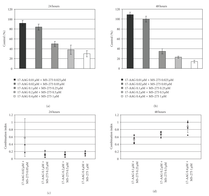
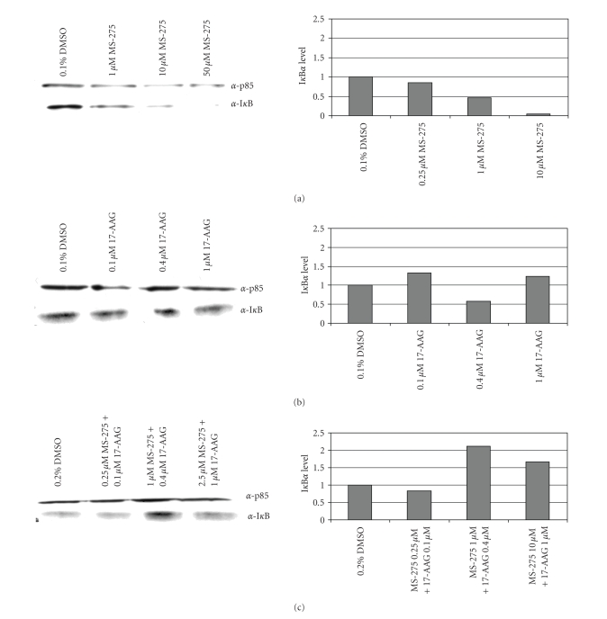
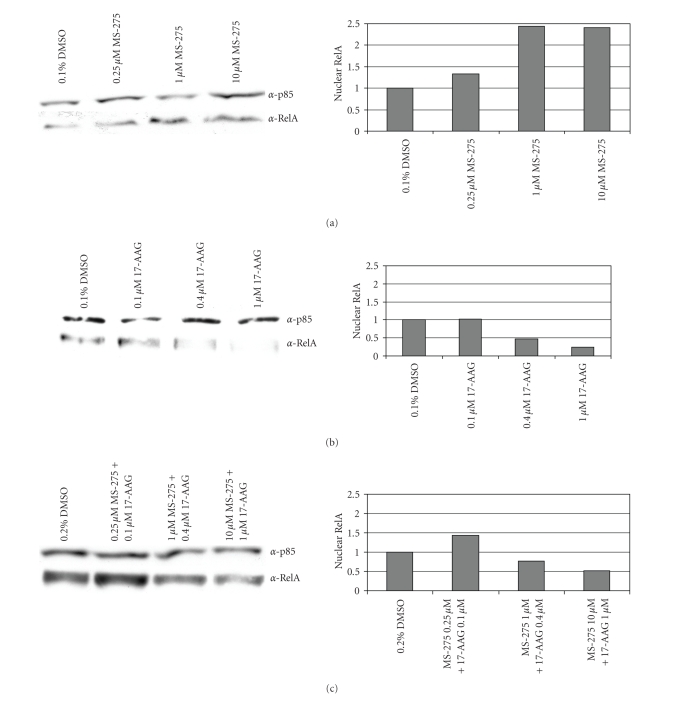
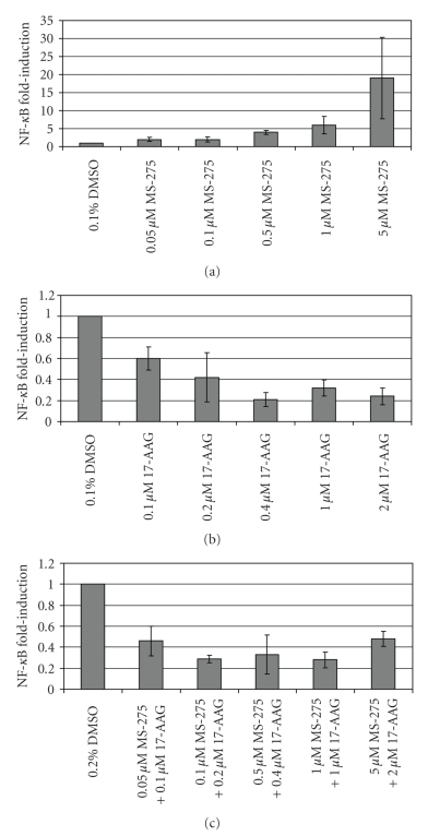
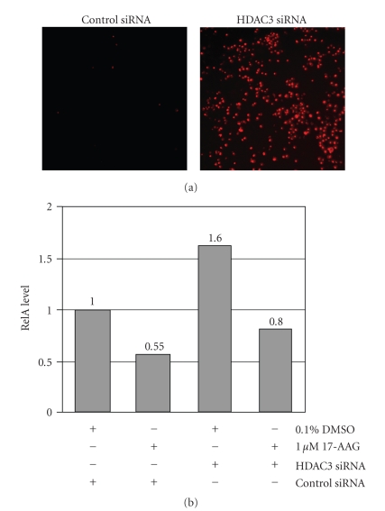
Current systemic therapies have little curative benefit for synovial sarcoma. Histone deacetylase (HDAC) inhibitors and the heat shock protein 90 (Hsp90) inhibitor 17-AAG have recently been shown to inhibit synovial sarcoma in preclinical models. We tested combinations of 17-AAG with the HDAC inhibitor MS-275 for synergism by proliferation and apoptosis assays. The combination was found to be synergistic at multiple time points in two synovial sarcoma cell lines. Previous studies have shown that HDAC inhibitors not only induce cell death but also activate the survival pathway NF-kappaB, potentially limiting therapeutic benefit. As 17-AAG inhibits activators of NF-kappaB, we tested if 17-AAG synergizes with MS-275 through abrogating NF-kappaB activation. In our assays, adding 17-AAG blocks NF-kappaB activation by MS-275 and siRNA directed against histone deacetylase 3 (HDAC3) recapitulates the effects of MS-275. Additionally, we find that the NF-kappaB inhibitor BAY 11-7085 synergizes with MS-275. We conclude that agents inhibiting NF-kappaB synergize with HDAC inhibitors against synovial sarcoma.


Figures

Figure 1
Figure 1
Effect of 17-AAG and MS-275 in combination on SYO-1 synovial sarcoma monolayer cultures. Cell survival at (a) 24 hours and (b) 48 hours for drugs combined at a set ratio of 2 parts 17-AAG to 5 parts MS-275. MTT cell proliferation assays were performed for the indicated doses, and survival is plotted relative to vehicle control. Corresponding combination indices are shown for (c) 24 and (d) 48 hours, where values below 1 indicate synergistic drug interactions. Error bars represent 95% confidence intervals. Combination indices were calculated using the Chou and Talalay Median Dose Method (not applicable at low doses where effective cell killing relative to control cell death was not observed).
Figure 2
Figure 2
Dose Response of IκBα. Immuunoblots and quantitation of IκBα levels after treatment with (a) MS-275, (b) 17-AAG and (c) Combination. Following 24 hour treatment total lysates were prepared and quantified. 10 μg of lysate was run on an SDS-page gel. By immunoblotting, IκBα and p85 (as a loading control) were detected using α-IκBα and α-p85 antibodies. Protein levels following treatment are compared to vehicle control which is set at 1.00.
Figure 3
Figure 3
Dose Response of RelA. Immunoblots and quantitation of nuclear RelA after treatment with (a) MS-275, (b ) 17-AAG and (c) Combination. Following 24 hour treatment, nuclear and cytoplasmic extracts were prepared and quantified. 15 μg were run on an SDS-page gel. Using immunoblotting techiniques, RelA and p85 (as a loading control) were detected using α-RelA and α-p85 antibodies. Protein levels following treatment are compared to vehicle control which is set at 1.00.
Figure 4
Figure 4
Transcriptional activation of NF-κB luciferase reporter. NF-κB induction following treatment with (a) MS-275, (b) 17-AAG, and (c) Combination. SYO-1 cells were grown as monolayer cultures in 24 well plates and transfected with 0.3 μg of NF-κB luciferase reporter plasmid. Cells were treated the following day for 24 hours and lysed with passive lysis buffer. Samples were aliquoted to plates and simultaneously assayed for luminosity by injection with 50 μl/well of LARII reagent and protein quantity by copper sulfate/bichionic acid assay. Readings for luminosity were normalized to protein concentration and vehicle control was set to 1.00. Error bars represent 95% confidence intervals for three replicate measurements.
Figure 5
Figure 5
HDAC3 siRNA knockdown recapitulates the effects HDAC inhibitors on synovial sarcoma cells. (a) 50 nM HDAC3 siRNA promotes extensive cell death at 24 hours, as shown by staining of a large inviable cell fraction by 500 ng/mL propidium iodide, where virtually all cells exposed to equimolar scrambled siRNA control are viable. (b) Densitometric quantitation of Western blots for nuclear RelA, expressed relative to control siRNA transfected cells treated with vehicle (1.0).

Similar articles

Cited by

References

    1. Ladanyi M. Fusions of the SYT and SSX genes in synovial sarcoma. Oncogene. 2001;20(40):5755–5762. - PubMed
    1. Ferrari A, Gronchi A, Casanova M, et al. Synovial sarcoma: a retrospective analysis of 271 patients of all ages treated at a single institution. Cancer. 2004;101(3):627–634. - PubMed
    1. Guillou L, Benhattar J, Bonichon F, et al. Histologic grade, but not SYT-SSX fusion type, is an important prognostic factor in patients with synovial sarcoma: a multicenter, retrospective analysis. Journal of Clinical Oncology. 2004;22(20):4040–4050. - PubMed
    1. Takenaka S, Ueda T, Naka N, et al. Prognostic implication of SYT-SSX fusion type in synovial sarcoma: a multi-institutional retrospective analysis in Japan. Oncology Reports. 2008;19(2):467–476. - PubMed
    1. Guadagnolo B, Zagars G, Ballo M, et al. Long-term outcomes for synovial sarcoma treated with conservation surgery and radiotherapy. International Journal of Radiation Oncology, Biology, Physics. 2007;69(4):1173–1180. - PubMed