Acute and late-onset optic atrophy due to a novel OPA1 mutation leading to a mitochondrial coupling defect
- PMID: 19325939
- PMCID: PMC2661005
Acute and late-onset optic atrophy due to a novel OPA1 mutation leading to a mitochondrial coupling defect
Abstract
Purpose: Autosomal dominant optic atrophy (ADOA, OMIM 165500), an inherited optic neuropathy that leads to retinal ganglion cell degeneration and reduced visual acuity during the early decades of life, is mainly associated with mutations in the OPA1 gene. Here we report a novel ADOA phenotype associated with a new pathogenic OPA1 gene mutation.
Methods: The patient, a 62-year-old woman, was referred for acute, painless, and severe visual loss in her right eye. Acute visual loss in her left eye occurred a year after initial presentation. MRI confirmed the diagnosis of isolated atrophic bilateral optic neuropathy. We performed DNA sequencing of the entire coding sequence and the exon/intron junctions of the OPA1 gene, and we searched for the mitochondrial DNA mutations responsible for Leber hereditary optic atrophy by sequencing entirely mitochondrial DNA. Mitochondrial respiratory chain complex activity and mitochondrial morphology were investigated in skin fibroblasts from the patient and controls.
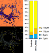
Results: We identified a novel heterozygous missense mutation (c.2794C>T) in exon 27 of the OPA1 gene, resulting in an amino acid change (p.R932C) in the protein. This mutation, which affects a highly conserved amino acids, has not been previously reported, and was absent in 400 control chromosomes. Mitochondrial DNA sequence analysis did not reveal any mutation associated with Leber hereditary optic neuropathy or any pathogenic mutations. The investigation of skin fibroblasts from the patient revealed a coupling defect of oxidative phosphorylation and a larger proportion of short mitochondria than in controls.
Conclusions: The presence of an OPA1 mutation indicates that this sporadic, late-onset acute case of optic neuropathy is related to ADOA and to a mitochondrial energetic defect. This suggests that the mutational screening of the OPA1 gene would be justified in atypical cases of optic nerve atrophy with no evident cause.
Figures


Similar articles
-
Genetic screening for OPA1 and OPA3 mutations in patients with suspected inherited optic neuropathies.Ophthalmology. 2011 Mar;118(3):558-63. doi: 10.1016/j.ophtha.2010.07.029. Epub 2010 Oct 30. Ophthalmology. 2011. PMID: 21036400 Free PMC article.
-
Novel mutations in the OPA1 gene and associated clinical features in Japanese patients with optic atrophy.Ophthalmology. 2006 Mar;113(3):483-488.e1. doi: 10.1016/j.ophtha.2005.10.054. Ophthalmology. 2006. PMID: 16513463
-
Fourteen novel OPA1 mutations in autosomal dominant optic atrophy including two de novo mutations in sporadic optic atrophy.Hum Mutat. 2003 Jun;21(6):656. doi: 10.1002/humu.9152. Hum Mutat. 2003. PMID: 14961560
-
OPA1-associated disorders: phenotypes and pathophysiology.Int J Biochem Cell Biol. 2009 Oct;41(10):1855-65. doi: 10.1016/j.biocel.2009.04.012. Epub 2009 Apr 21. Int J Biochem Cell Biol. 2009. PMID: 19389487 Review.
-
Recessive optic atrophy, sensorimotor neuropathy and cataract associated with novel compound heterozygous mutations in OPA1.Mol Med Rep. 2016 Jul;14(1):33-40. doi: 10.3892/mmr.2016.5209. Epub 2016 May 4. Mol Med Rep. 2016. PMID: 27150940 Free PMC article. Review.
Cited by
-
Whole-Exome Sequencing Identifies Novel Variants that Co-segregates with Autosomal Recessive Retinal Degeneration in a Pakistani Pedigree.Adv Exp Med Biol. 2018;1074:219-228. doi: 10.1007/978-3-319-75402-4_27. Adv Exp Med Biol. 2018. PMID: 29721947 Free PMC article.
-
Genetic screening for OPA1 and OPA3 mutations in patients with suspected inherited optic neuropathies.Ophthalmology. 2011 Mar;118(3):558-63. doi: 10.1016/j.ophtha.2010.07.029. Epub 2010 Oct 30. Ophthalmology. 2011. PMID: 21036400 Free PMC article.
-
[Hereditary optic atrophies].Ophthalmologe. 2009 Sep;106(9):845-57. doi: 10.1007/s00347-009-2023-0. Ophthalmologe. 2009. PMID: 19756638 German.
-
Dominant Optic Atrophy (DOA): Modeling the Kaleidoscopic Roles of OPA1 in Mitochondrial Homeostasis.Front Neurol. 2021 Jun 9;12:681326. doi: 10.3389/fneur.2021.681326. eCollection 2021. Front Neurol. 2021. PMID: 34177786 Free PMC article. Review.
-
Metabolically induced heteroplasmy shifting and l-arginine treatment reduce the energetic defect in a neuronal-like model of MELAS.Biochim Biophys Acta. 2012 Jun;1822(6):1019-29. doi: 10.1016/j.bbadis.2012.01.010. Epub 2012 Jan 28. Biochim Biophys Acta. 2012. PMID: 22306605 Free PMC article.
References
-
- Johnston PB, Gaster RN, Smith VC, Tripathi RC. A clinico-pathological study of autosomal dominant optic atrophy. Am J Ophthalmol. 1979;88:868–75. - PubMed
-
- Hoyt CS. Autosomal dominant optic atrophy: a spectrum of disability. Ophthalmology. 1980;87:245–51. - PubMed
-
- Lyle WM. Genetic risks. Waterloo, Ontario: Univ of Waterloo Press;1990.
-
- Kjer B, Eiberg H, Kjer P, Rosenberg T. Dominant optic atrophy mapped to chromosome 3q region II. Clinical and epidemiological aspects. Acta Ophthalmol Scand. 1996;74:3–7. - PubMed
-
- Votruba M, Fitzke FW, Holder GE, Carter A, Bhattacharya SS, Moore AT. Clinical features in affected individuals from 21 pedigrees with dominant optic atrophy. Arch Ophthalmol. 1998;116:351–8. - PubMed
Publication types
MeSH terms
Substances
LinkOut - more resources
Full Text Sources
Medical
Molecular Biology Databases
