. 2009 Sep-Oct;11(5):343-55.
doi: 10.1007/s11307-009-0215-2. Epub 2009 Mar 27.

NCI-sponsored trial for the evaluation of safety and preliminary efficacy of 3'-deoxy-3'-[18F]fluorothymidine (FLT) as a marker of proliferation in patients with recurrent gliomas: preliminary efficacy studies

Affiliations

NCI-sponsored trial for the evaluation of safety and preliminary efficacy of 3'-deoxy-3'-[18F]fluorothymidine (FLT) as a marker of proliferation in patients with recurrent gliomas: preliminary efficacy studies

Alexander M Spence et al. Mol Imaging Biol. 2009 Sep-Oct.

Abstract

Purpose: 3'-Deoxy-3'-[18F]fluorothymidine ([18F]FLT) is being developed for imaging cellular proliferation. The goals were to explore the capacity of FLT-positron emission tomography (PET) to distinguish between recurrence and radionecrosis in gliomas and compare the results to those obtained with 2-fluoro-2-deoxy-D: -glucose (FDG).

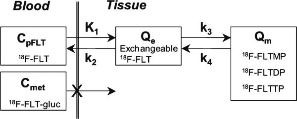
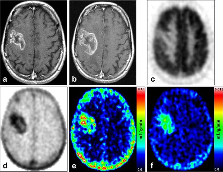
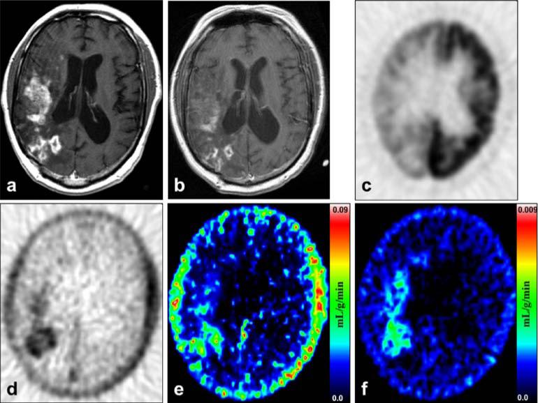
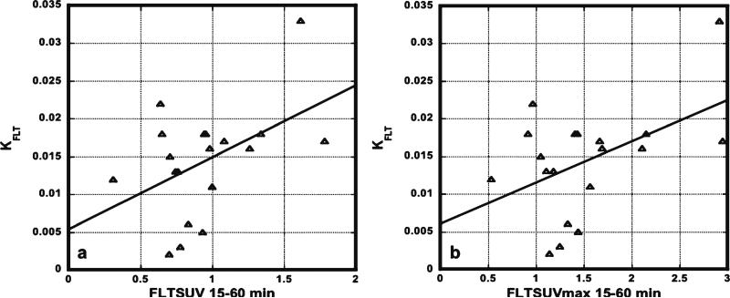
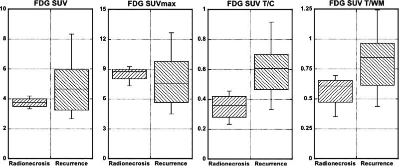
Procedures: Fifteen patients with tumor recurrence and four with radionecrosis, determined by clinical course and magnetic resonance imaging results, were studied by dynamic [18F]FLT-PET with arterial blood sampling. A two-tissue compartment four-rate constant model was used to determine metabolic flux (K (FLT)), blood to tissue transport (K (1)), and phosphorylation (k (3)). FDG-PET scans were obtained 75-90 min postinjection.

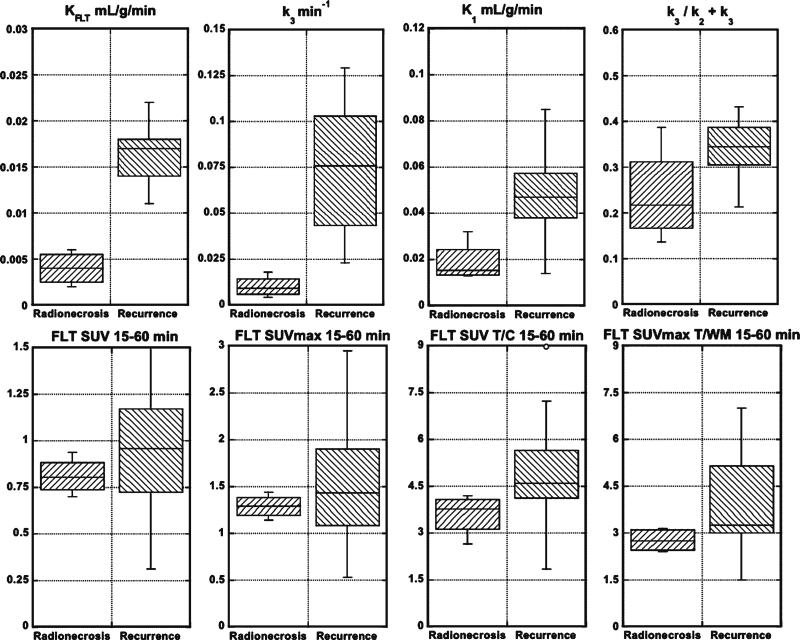
Results: K (FLT) and k (3), but not K (1) or k (3)/k (2) + k (3), reached significance for separating the recurrence from radionecrosis groups. Standardized uptake value and visual analyses of FLT or FDG images did not reach significance.

Conclusions: K (FLT) (flux) appears to distinguish recurrence from radionecrosis better than other parameters, FLT and FDG semiquantitative approaches, or visual analysis of images of either tracer.


Figures

Fig. 1
Fig. 1
The kinetic model of FLT metabolism is comprised of an exchangeable tissue compartment (Qe) and a compartment of trapped FLT nucleotides (Qm). Four rate constants (K1k4) describe the kinetic transfer rates between the two compartments and blood. FLTMP FLT-monophosphate, FLTDP FLT-diphosphate, FLTTP FLT-triphosphate, FLT-gluc FLT-glucuronide synthesized in the liver, CpFLT concentration of FLT in arterial plasma, Cmet concentration of metabolites in arterial plasma.
Fig. 2
Fig. 2
Recurrence: Patient 17 was a 41-year-old woman who died 6 months after the FLT-PET study from progression of her recurrent glioblastoma multiforme. a MRI T1+Gd shows a large right frontal contrast-enhancing lesion at the time of her FLT-PET and FDG-PET scans. b MRI T1+Gd taken 2.5 months after the FLT-PET shows definite progression of the contrast enhancing disease. c FDG-PET of the same plane and time as in a shows that the tumor has uptake focally less than cortex but greater than white matter. d FLT-PET shows a high level of uptake corresponding to the MRI T1+Gd abnormality in a. e Mixture analysis image of the K1 transport parameter. f Mixture analysis image of the KFLT flux parameter. Note that the color scale is ten times less for f than for e.
Fig. 3
Fig. 3
Radionecrosis: Patient 1, a 46-year-old man with a right temporal grade 2 oligodendroglioma diagnosed by biopsy 9 years before FLT-PET and treated with radiotherapy 53 months before FLT-PET. a MRI T1+Gd shows extensive contrast enhancement in the right temporal, frontal, and parietal lobes. b MRI T1+Gd obtained 13 months after the FLT-PET shows decidedly less volume of enhancement. c FDG-PET of the same plane and time as a shows that the tracer uptake is focally greater than white matter but less than cortex. d FLT-PET shows a high level of uptake corresponding closely to the MRI T1+Gd abnormality in a. e Mixture analysis image of the K1 transport parameter. f Mixture analysis image of the KFLT flux parameter. Note that the color scale is ten times less for f than for e.
Fig. 4
Fig. 4
Box plots showing the comparisons between the radionecrosis and recurrence groups for KFLT, k3, K1, k3/k2+k3, FLT SUV 15–60 min, FLT SUVmax 15–60 min, FLT SUV T/C 15–60 min, and FLT SUVmax T/WM 15–60 min. See Table 3 for p values.
Fig. 5
Fig. 5
Correlations between KFLT and SUV 15–60 (a) or SUVmax 15–60 (b) were poor (n=19). R2 values were only 0.22 and 0.23, respectively.
Fig. 6
Fig. 6
Box plots showing the comparisons between the radionecrosis and recurrence groups for FDG SUV, FDG SUVmax, FDG SUV T/C, and FDG SUV T/WM. See Table 5 for p values.

Similar articles

Cited by

References

    1. Grierson JR, Schwartz JL, Muzi M, Jordan R, Krohn KA. Metabolism of 3′-deoxy-3′-[F-18]fluorothymidine in proliferating A549 cells: validations for positron emission tomography. Nucl Med Biol. 2004;31:829–837. - PubMed
    1. Brandes AA, Tosoni A, Spagnolli F, et al. Disease progression or pseudoprogression after concomitant radiochemotherapy treatment: pitfalls in neurooncology. Neuro Oncol. 2008;10:361–367. - PMC - PubMed
    1. Chamberlain MC, Glantz MJ, Chalmers L, Van Horn A, Sloan AE. Early necrosis following concurrent Temodar and radiotherapy in patients with glioblastoma. J Neurooncol. 2007;82:81–83. - PubMed
    1. Spence AM, Muzi M, Link JM, Hoffman JM, Eary JF, Krohn KA. NCI-sponsored trial for the evaluation of safety and preliminary efficacy of FLT as a marker of proliferation in patients with recurrent gliomas: safety studies. Mol Imaging Biol. 2008;10:271–280. - PMC - PubMed
    1. Muzi M, Spence AM, O'Sullivan F, et al. Kinetic analysis of 3′-deoxy-3′-18F-fluorothymidine in patients with gliomas. J Nucl Med. 2006;47:1612–1621. - PubMed

Publication types

MeSH terms