. 2009 Jun 1;514(4):297-309.
doi: 10.1002/cne.22022.

Human neural stem cell grafts in the spinal cord of SOD1 transgenic rats: differentiation and structural integration into the segmental motor circuitry

Affiliations

Human neural stem cell grafts in the spinal cord of SOD1 transgenic rats: differentiation and structural integration into the segmental motor circuitry

Leyan Xu et al. J Comp Neurol. .

Abstract

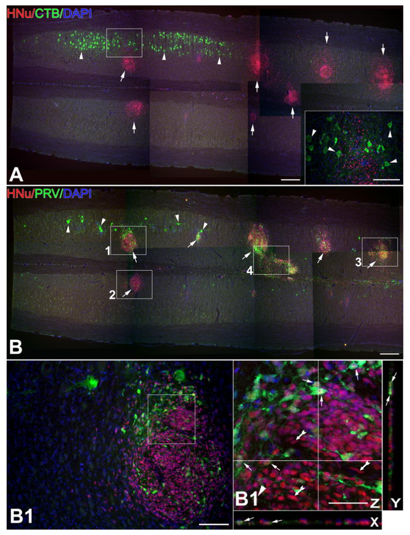
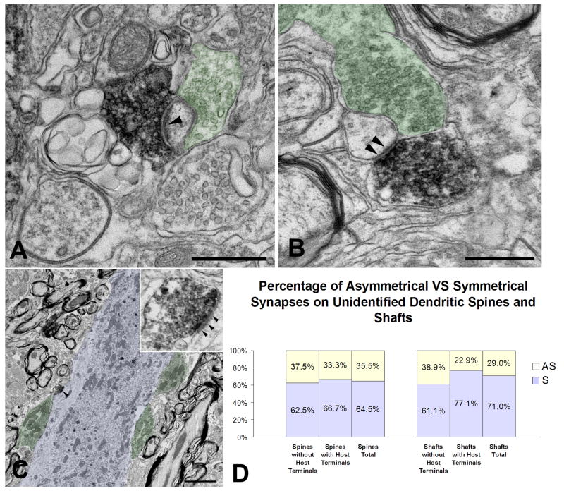
Cell replacement strategies for degenerative and traumatic diseases of the nervous system depend on the functional integration of grafted cells into host neural circuitry, a condition necessary for the propagation of physiological signals and, perhaps, targeting of trophic support to injured neurons. We have recently shown that human neural stem cell (NSC) grafts ameliorate motor neuron disease in SOD1 transgenic rodents. Here we study structural aspects of integration of neuronally differentiated human NSCs in the motor circuitry of SOD1 G93A rats. Human NSCs were grafted into the lumbar protuberance of 8-week-old SOD1 G93A rats; the results were compared to those on control Sprague-Dawley rats. Using pre-embedding immuno-electron microscopy, we found human synaptophysin (+) terminals contacting the perikarya and proximal dendrites of host alpha motor neurons. Synaptophysin (+) terminals had well-formed synaptic vesicles and were associated with membrane specializations primarily in the form of symmetrical synapses. To analyze the anatomy of motor circuits engaging differentiated NSCs, we injected the retrograde transneuronal tracer Bartha-pseudorabies virus (PRV) or the retrograde marker cholera toxin B (CTB) into the gastrocnemius muscle/sciatic nerve of SOD1 rats before disease onset and also into control rats. With this tracing, NSC-derived neurons were labeled with PRV but not CTB, a pattern suggesting that PRV entered NSC-derived neurons via transneuronal transfer from host motor neurons but not via direct transport from the host musculature. Our results indicate an advanced degree of structural integration, via functional synapses, of differentiated human NSCs into the segmental motor circuitry of SOD1-G93A rats.


Figures

Figure 1
Figure 1
Synaptic contacts between human NSC- derived neurons and host motor neurons in SOD1 G93A rats, as shown with immunofluorescence (A-A′) and immuno-electron microscopy (B-E). In all cases, human synaptophysin immunoreactivity serves as a marker for NSC-derived terminals. Postsynaptic host structures are labeled with phenotypic markers such as choline acetyltransferase (ChAT) in (A) or left unlabeled as in (B-E). The host (rat) identity of postsynaptic structure in Fig. 1 derives from the classical motor neuron morphology of post-synaptic perikarya and the fact that no human NSCs differentiate into motor neurons in these experiments. A-A′. A large number of human synaptophysin (+) boutons (SYN, red; arrows) contact host ChAT (+) motor neurons (green). A′ is a confocal image taken from the same section as in (A) to confirm the apposition of human synaptophysin (+) boutons to the cell body and dendrites of the large motor neuron on the right of (A). Arrows in (A′) depict boutons that are further validated with x and y resectioning. B-D. These electron photomicrographs depict, in successive enlargements, a host motor neuron (delineated with a black dashed line in [B]) contacted by a human synaptophysin immunoreactive terminal (arrow in all panels); the latter stands in comparison to an adjacent, unlabeled, host terminal (double arrow head in panels [B-C]). Panel D shows the ultrastructure of the human NSC-derived terminal replete with round synaptic boutons, mitochondria and terminal membrane specializations (arrowheads). E. This photograph is taken from a section immediately adjacent to the one in (B-D) and showcases the same human NSC-derived synapse as panels B-D (arrow) and, in greater detail, the adjacent host-derived (unlabeled) terminal (double arrow head). Magenta-green color copy of Figure 1 is also available as Supplementary Figure 1. Size bars: A, 50μm; A′, 20 μm; B, 10 μm; C, 1 μm; D, 0.1 μm; E, 0.5 μm
Figure 2
Figure 2
Circuits engaging differentiated NSCs in SOD1 G93A rat spinal cord, as demonstrated with CBT and PRV tracing from injections into the gastrocnemius/sciatic nerve. Panels with letters-numbers represent sequential magnifications of framed areas in (B). A-B. These two panels are taken from adjacent sections treated with CTB (A) and PRV (B) ICC and are juxtaposed to demonstrate differential patterns of labeling. In (A), note the selective CTB labeling of large motor neurons in the dorsolateral column (arrowheads) ipsilateral to the right-hand injection side and absence of any labeling in the graft (arrows pointing to HNu [red]-labeled structures). In contrast, there is sparse PRV labeling of motor neurons (asterisk) on the side of tracer injection and an especially intense labeling of graft cells (arrows) on both sides in (B). B1-B2′. These panels represent serial magnifications of framed areas in B and B1-2, such that (B1′) is a magnification of the framed area in (B1) and so is (B2′) with respect to (B2). Panel B1 showcases a PRV-labeled host motor neuron (asterisk) and isolated as well as clustered NSCs labeled with PRV (arrows). Panel B1′ is a confocal image showing multiple NSC-derived, PRV-labeled neurons (arrows; dually fluorescent for HNu-red and PRV-green) and many PRV (-) human NSCs (tailed arrows). Panel B2′ is a confocal image from a region where PRV-immunoreactive host neurons (arrowheads), non PRV-labeled human NSCs (tailed arrows) mingled with PRV- and HNu-labeled graft-derived neurons (arrows). C. This cresyl-violet stained section is adjacent to sections depicted in (A-B) and serves as a guide for spinal cord anatomy including graft sites (GR) and ventral horn (VH). Note that motor neuron degeneration is not evident at this time point in the natural course of G93A motor neuron disease. D. NSC-derived neurons (here dually labeled with HNu-red and PRV-green) in lamina X ventral to the central canal were consistently present in PRV-stained material and may have played a role for transneuronal transfer or PRV from side to side. A PRV (+) host neuron is indicated with an arrowhead, whereas a PRV (+) graft-derived neurons (also positive for HNu) is shown with an arrow. Magenta-green color copy of Figure 2 is also available as Supplementary Figure 2. Size bars: A, 200 μm; B, 200 μm; B1, 50 μm; B′, 10 μm; B2, 50 μm; B2′, 10 μm; C, 200 μm; D, 10 μm
Figure 3
Figure 3
Circuits engaging differentiated NSCs in the normal spinal cord of normal SD rats as demonstrated with CBT (A) and PRV (B) tracing from injections into the gastrocnemius/sciatic nerve. Panels with letters-numbers represent sequential magnifications of framed areas in (B). A-B. These two panels are taken from adjacent longitudinal sections processed with ICC for CTB (A) and PRV (B) visualization and juxtaposed here to reveal differential patterns of labeling. (A) features the selective CTB labeling of α-motor neurons in the dorsolateral column (arrowheads) ipsilateral to the side of tracer injection and the absence of any labeling in the graft (shown here with arrows as HNu [red]-labeled structures). PRV preparations (B), in contrast, show sparse labeling of motor neurons (arrowheads) on the injection side and intense labeling of cells in grafts (arrows). Inset represents magnification of framed area in main panel and shows a region where CTB-labeled host motor neurons (arrowheads) mingle with unlabeled, HNu (+), graft-derived cells. B1-B4′. The four main panels (B1-B4) are magnifications of framed areas in (B) and panels further designated with a prime sign are confocal magnifications of main panels to showcase PRV-labeled, graft-derived (HNu [+]) neurons within grafts. (B2) is from the side contralateral to PRV injection. (B4) depicts a bridge between two HNu (+) inoculation sites with fusiform cells that may migrate from one site to another. Double-labeled (graft-derived) profiles are indicated with arrows and PRV (-) human NSCs are indicated with tailed arrows in all panels. Magenta-green color copy of Figure 3 is also available as Supplementary Figure 3. Size bars: A, 200 μm; B, 200 μm; B1, 50 μm; B1′, 10 μm; B2, 50 μm; B2′, 10 μm; B3, 50 μm; B3′, 10 μm; B4, 50 μm; B4′, 10 μm
Figure 4
Figure 4
Representative synaptic profiles of graft-derived, human synaptophysin (+) boutons contacting dendritic shafts and spines of identified host motor neurons. Panels labeled with a primed capital letter represent magnifications of framed areas in panels labeled with capital letter alone. Green and blue colors denote host pre- and post-synaptic structures, respectively. A-A′. This EM photograph shows a human synaptophysin (+) terminal swelling (arrowheads) forming an axodendritic symmetrical synapse with the dendritic shaft of a host motor neuron; host synaptic terminals are also shown on the same dendrite. B. A graft-derived, human synaptophysin (+) terminal squeezes in to form an axodendritic synapse (arrowheads) on a dendritic shaft traceable to a host motor neuron, along with 8 host-derived synapses (green). Inset represents a higher magnification of the human synaptophysin (+) terminal in main panel. C-C′. A graft-derived terminal forms a symmetrical axospinous synapse (arrowhead) on the proximal dendrite of a host motor neuron. Inset in C′ represents a higher magnification of the labeled synapse in main panel. Spines traceable to host motor neurons are rare in our preparations. D. This panel shows a graft-derived en passant swelling forming an asymmetrical synapse (arrowhead) with a host motor neuron cell body; inset shows detail of the synapse in higher magnification. E. A graft-derived, human synaptophysin (+) terminal forms an axoaxonal synapse (arrowheads) on a host terminal that, in turn, synapses on to a host motor neuron. F. A bar diagram showing percentage rates of asymmetrical (AS) versus symmetrical (S) graft derived, human synaptophysin (+) synapses on identified motor neuron postsynaptic (cell bodies and dendritic shaft; spines traceable to motor neurons are very rare). Size bars: A, 2 μm; A1′, 0.5 μm; B, 1 μm; C, 20 μm; C′, 2 μm; D, 0.5 μm; E, 0.5 μm
Figure 5
Figure 5
Representative synaptic profiles of graft-derived terminals contacting dendritic shafts and spines in the host ventral horn that could not be traced to specific neuron types, such as motor neurons. Some of these postsynaptic structures also receive innervation by human synaptophysin (-) (host) terminals. All profiles accounted for here are far from the graft inoculation site. As in Fig. 4, green and blue colors denote host pre- and post-synaptic structures, respectively. A-B. These two panels show graft-derived terminals forming asymmetrical (A) and symmetrical (B) axospinous synapses (arrowheads) on unidentified dendritic spines that also receive synaptic contacts from host neurons. C. A graft-derived, human synaptophysin (+) terminal forms an asymmetrical synapse (arrowhead) with unidentified dendritic shaft. Host terminals are also shown on the same portion of the host dendrite. Inset shows further detail of the graft-to-host synapse in the main panel. D. This bar diagram shows the percentage rates of asymmetrical (AS) versus symmetrical (S) graft derived, human synaptophysin (+) synapses on unidentified dendritic shafts and spines, further subdivided per the co-occurrence or not of host synapses in the vicinity. Size bars: A, 0.5 μm; B, 0.5 μm; C, 2 μm

References

    1. Akiyama Y, Honmou O, Kato T, Uede T, Hashi K, Kocsis JD. Transplantation of clonal neural precursor cells derived from adult human brain establishes functional peripheral myelin in the rat spinal cord. Exp Neurol. 2001;167:27–39. - PubMed
    1. Anderson L, Caldwell MA. Human neural progenitor cell transplants into the subthalamic nucleus lead to functional recovery in a rat model of Parkinson's disease. Neurobiol Dis. 2007;27:133–140. - PubMed
    1. Aoki M, Kato S, Nagai M, Itoyama Y. Development of a rat model of amyotrophic lateral sclerosis expressing a human SOD1 transgene. Neuropathology. 2005;25:365–370. - PubMed
    1. Aston-Jones G, Card JP. Use of pseudorabies virus to delineate multisynaptic circuits in brain: opportunities and limitations. J Neurosci Methods. 2000;103:51–61. - PubMed
    1. Bareyre FM, Kerschensteiner M, Raineteau O, Mettenleiter TC, Weinmann O, Schwab ME. The injured spinal cord spontaneously forms a new intraspinal circuit in adult rats. Nat Neurosci. 2004;7:269–277. - PubMed

Publication types

MeSH terms