. 2009 Sep;18(7):955-68.
doi: 10.1089/scd.2008.0310.

Phenotypic characterization, osteoblastic differentiation, and bone regeneration capacity of human embryonic stem cell-derived mesenchymal stem cells

Affiliations

Phenotypic characterization, osteoblastic differentiation, and bone regeneration capacity of human embryonic stem cell-derived mesenchymal stem cells

Premjit Arpornmaeklong et al. Stem Cells Dev. 2009 Sep.

Abstract

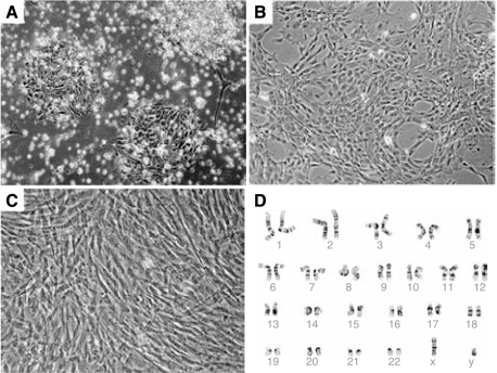
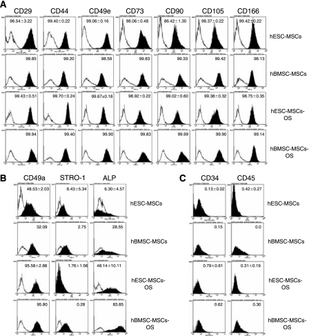
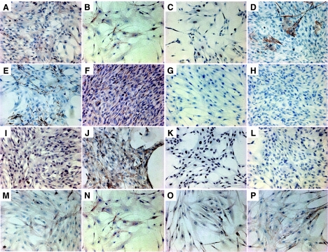
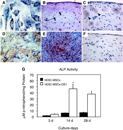
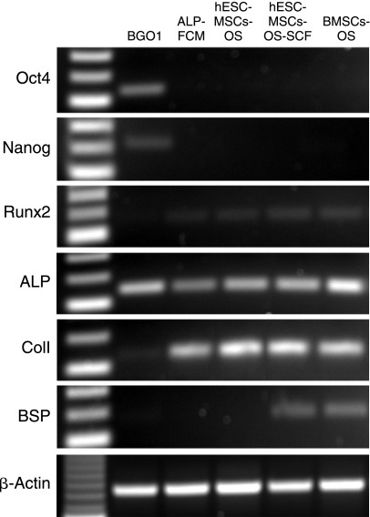
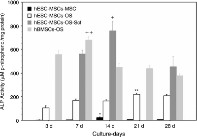
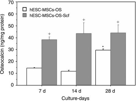
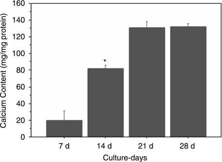
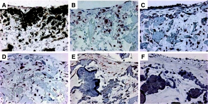
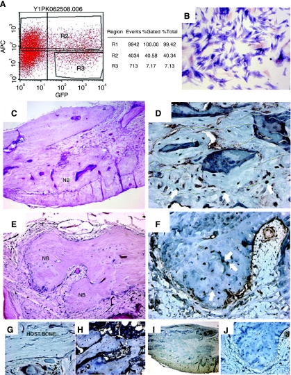
To enhance the understanding of differentiation patterns and bone formation capacity of hESCs, we determined (1) the temporal pattern of osteoblastic differentiation of human embryonic stem cell-derived mesenchymal stem cells (hESC-MSCs), (2) the influence of a three-dimensional matrix on the osteogenic differentiation of hESC-MSCs in long-term culture, and (3) the bone-forming capacity of osteoblast-like cells derived from hESC-MSCs in calvarial defects. Incubation of hESC-MSCs in osteogenic medium induced osteoblastic differentiation of hESC-MSCs into mature osteoblasts in a similar chronological pattern to human bone marrow stromal cells and primary osteoblasts. Osteogenic differentiation was enhanced by culturing the cells on three-dimensional collagen scaffolds. Fluorescent-activated cell sorting of alkaline phosphatase expressing cells was used to obtain an enriched osteogenic cell population for in vivo transplantation. The identification of green fluorescence protein and expression of human-specific nuclear antigen in osteocytes in newly formed bone verified the role of transplanted human cells in the bone regeneration process. The current cell culture model and osteogenic cell enrichment method could provide large numbers of osteoprogenitor cells for analysis of differentiation patterns and cell transplantation to regenerate skeletal defects.


Figures

FIG. 1.
FIG. 1.
Phase contrast images demonstrating morphological changes in aggregates of human embryonic stem cells (hESCs) cultured in medium supplemented with 10% FBS (20×): (A) 2 days after initial cell seeding; (B) after the first cell passage; (C) after the seventh cell passage; and (D) a normal karyotype for hESC mesenchymal stem cells eight passages after derivation.
FIG. 2.
FIG. 2.
Surface antigen profiling of human embryonic stem cell–derived mesenchymal stem cells (hESC-MSCs) and human bone marrow stromal cells (hBMSCs) in MSC and osteogenic medium (OS). Cells were labeled and fluorescent-activated cell sorted (FACS) with: (A) antibodies against the MSC surface antigens that exhibited expression levels greater than 95%: CD29-FITC, CD44-APC, CD49e-PE, CD73-PE, CD90-PE-CY5™, CD105-PE, and CD166-PE; (B) antibodies against surface antigens that either increased or decreased markedly in osteogenic medium: CD49a-PE, STRO-1-PE, and ALP-APC; and (C) hematopoietic stem cell surface antigens: CD34 PE-CY5™ and CD45-FITC. Data from hESC-MSCs and hESC-MSCs-OS were average values from two different cell strains at passage 6 and data from hBMSC and hBMSC-OS were derived from analysis of cells from one cell strain at passage 4. The white histogram represents isotype controls and the black histogram represents the conjugated antibody of each antigen.
FIG. 3.
FIG. 3.
Immunohistochemical staining of surface antigens on human embryonic stem cell–derived mesenchymal stem cells (hESC-MSCs) cultured in MSC medium (A–J), hESC-MSCs in osteogenic medium (K), negative control (L), and human bone marrow stromal cells (hBMSCs) in MSC medium (MP). Positive staining is indicated by red-brown cytoplasmic staining in all images except nuclear staining of brachyury (C) and Runx2 (K). (A) hESC-MSCs-CD105, (B) CD166, (C) brachyury, (D) smooth muscle α-actin, (E) collagen type I, (F) vimentin, (G) VCAM-1, (H) α-fetoprotein, (I) estrogen receptor α (ERα), and (J) β-catenin. (K) hESC-MSCs in osteogenic medium-Runx2. (L) Negative control omitting monoclonal antibody. (M) hBMSC-MSCs-CD105, (N) CD166, (O) ERα, and (P) β-catenin. (AEC substrate and hematoxylin counter stain, magnification 20×.)
FIG. 4.
FIG. 4.
Phase contrast images of functional differentiation of human embryonic stem cell–derived mesenchymal stem cells (hESC-MSCs) in (A) adipogenic medium with Oil Red O staining on Day 14 demonstrating red lipid droplets in cytoplasm (20×); (B) chondrogenic medium with safranin O staining of the extracellular matrix (ECM) and cells with large nuclei in lacunae (arrow) on Day 21 (40×); (C) immunohistochemical staining showing patchy red staining of collagen type II (arrowheads); (D) Alkaline phosphatase (ALP) staining (blue staining) in hESC-MSCs in osteogenic medium (OS1) with neutral red counter stain on Day 14; (E) alizarin red staining of mineralized ECM on Day 21 (20×); (F) negative control with omission of a primary antibody; (G) Progression of ALP activity of hESC-MSCs in MSC medium, hESC-MSCs, and hESC-MSCs in osteo-genic medium, hESC-MSCs-OS1, for a period of 28 days. The symbol (*) represents the highest level of ALP activity in comparison within and between groups (P < 0.01) (n = 5).
FIG. 5.
FIG. 5.
Gene transcription analysis comparing expression of pluripotent regulator genes, Oct4 and Nanog and the osteoblast-associated genes, Runx2, alkaline phosphatase (ALP), collagen type I (ColI), and bone sialoprotein (BSP) in undifferentiated hESCs at passage 58 (lane 1, BGO1), FCM-sorted ALP expressing cells (lane 2, ALP-FCM), hESC-MSCs in osteogenic medium (lane 3, hESC-MSCs-OS), on three-dimensional collagen scaffolds (lane 4, hESC-MSCs-OS-Scf), and BMSCs in osteogenic medium (lane 5, BMSCs-OS) on culture-day 14. BMSCs-OS served as positive control and β-actin as a housekeeping gene. See Table 1 for primers and amplicon sizes.
FIG. 6.
FIG. 6.
Alkaline phosphatase (ALP) activity in cultured cells. Human embryonic stem cell–derived mesenchymal stem cells (hESC-MSCs) in MSC medium (hESC-MSCs-MSC); hESC-MSCs in osteogenic medium (hESC-MSCs-OS); hESC-MSCs in osteogenic medium on collagen scaffolds (hESC-MSCs-OS-Scf); and human bone marrow stromal cells (hBMSCs) in osteogenic medium (hBM-SCs-OS), for a period of 28 days. The symbol (*) represents significant changes in ALP activity of hESC-MSCs (P < 0.01), (**) hESC-MSCs-OS (P < 0.01), (+) hESC-MSCs-OS-Scf (P < 0.05), and (++) hBMSCs-OS (P < 0.01). n = 5, mean ± SEM.
FIG. 7.
FIG. 7.
Levels of osteocalcin secretion in cultured cells. Human embryonic stem cell–derived mesenchymal stem cells (hESC-MSCs) in osteogenic medium (OS) on cell culture plates and on collagen scaffolds (Scf) for 28 days. The symbol (*) represents a significant increase in osteocalcin secretion from Days 7 and 14 to Day 28 (P < 0.05) and (+) significant higher levels of osteocalcin in hESC-MSCs-OS-Scf than hESC-MSCs-OS (P < 0.05) (n = 4, mean ± SEM).
FIG. 8.
FIG. 8.
Calcium content in the extracellular matrix (ECM) of human embryonic stem cell–derived mesenchymal stem cells (hESC-MSCs) in osteogenic medium on cell culture plates for a culture period for 28 days. The symbol (*) represents significant difference from Days 7 and 21 and 28 (P < 0.01) (n = 4, mean ± SEM).
FIG. 9.
FIG. 9.
von Kossa and immunohistochemical staining of paraffin-embedded sections of human embryonic stem cell–derived mesenchymal stem cells (hESC-MSCs) in osteogenic medium on collagen scaffold for 28 days demonstrating mineralization of the extracellular matrix (A) and deposition of noncollagenous bone matrix on the scaffold (BE) (20X). (A) von Kossa staining (black stain) and immunohistochemical staining of noncollagenous matrix proteins (B) ALP, (C) osteonectin, (D) osteopontin, and (E) osteocalcin secreted by cells on the scaffold. (F) Negative control with omission of primary antibodies. The antigen–antibody complexes were detected by AEC substrate yielding a red-brown stain (BE).
FIG. 10.
FIG. 10.
Intramembranous bone formation in calvarial defects by the transplanted osteoblast-like cells derived from human embryonic stem cell–derived mesenchymal stem cells (hESC-MSCs) cultured in osteogenic medium. (A) FAC sorting of APC-conjugated alkaline phosphatase (ALP) and green fluorescent protein (GFP) of cells before transplantation (Gate R2). (B) ALP staining of the sorted ALP-positive cells (blue) counter-stained with neutral red (red) (20×). (C,E) Histological images of newly formed bone (NB) near the margins of the defect (H&E, 40×). (D) Staining of human-specific nuclear antigen (HuNu) and (F) GFP staining of osteocytes in new bone matrix (arrow). (G) Negative staining of HuNu mAb on the mature host bone. (H) HuNu staining of mouse femur. (I) Negative control of HuNu and (J) GFP staining with omission of primary antibodies. Magnification of images CF was 40× and GJ was 20×.

References

    1. Mimeault M. Hauke R. Batra SK. Stem cells: a revolution in therapeutics-recent advances in stem cell biology and their therapeutic applications in regenerative medicine and cancer therapies. Clin Pharmacol Ther. 2007;82:252–264. - PubMed
    1. Kwan MD. Slater BJ. Wan DC. Longaker MT. Cell-based therapies for skeletal regenerative medicine. Hum Mol Genet. 2008;17:R93–R98. - PubMed
    1. Carpenter MK. Rosler E. Rao MS. Characterization and differentiation of human embryonic stem cells. Cloning Stem Cells. 2003;5:79–88. - PubMed
    1. Fenno LE. Ptaszek LM. Cowan CA. Human embryonic stem cells: emerging technologies and practical applications. Curr Opin Genet Dev. 2008;18:324–329. - PubMed
    1. Olivier EN. Rybicki AC. Bouhassira EE. Differentiation of human embryonic stem cells into bipotent mesenchymal stem cells. Stem Cells. 2006;24:1914–1922. - PubMed

Publication types

Substances