. 2009 Jul 1;315(11):1875-88.
doi: 10.1016/j.yexcr.2009.03.010. Epub 2009 Mar 24.

Anchorage-independent growth of breast carcinoma cells is mediated by serum exosomes

Affiliations

Anchorage-independent growth of breast carcinoma cells is mediated by serum exosomes

Josiah Ochieng et al. Exp Cell Res. .

Abstract

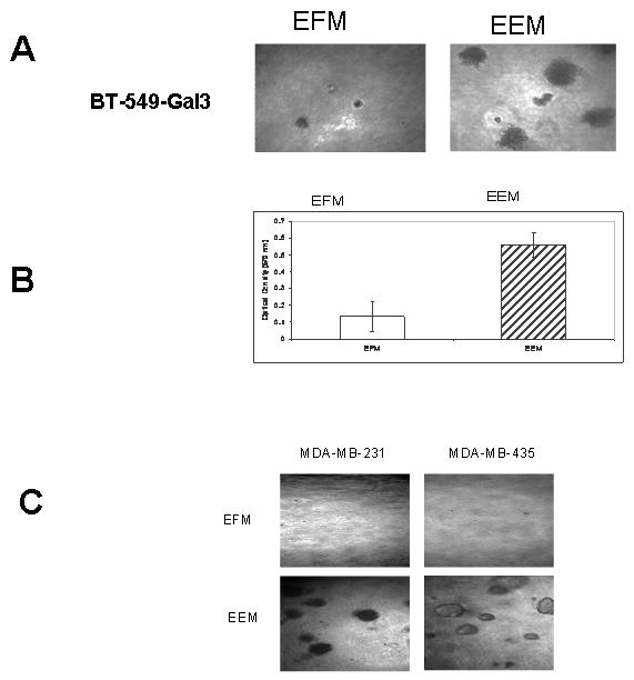
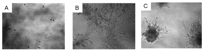
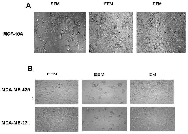
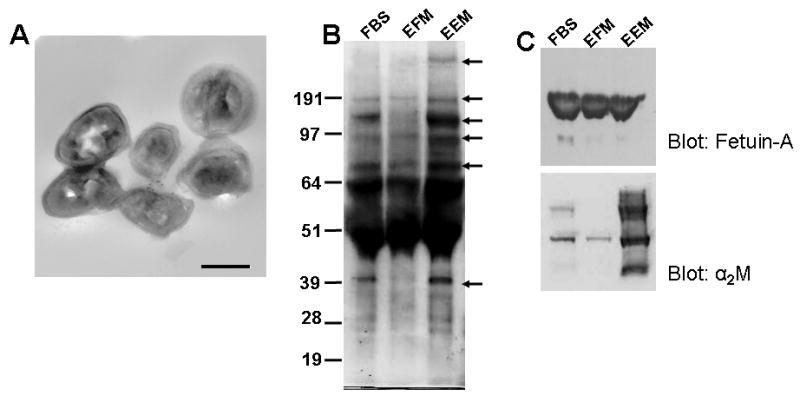
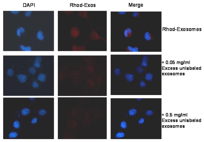
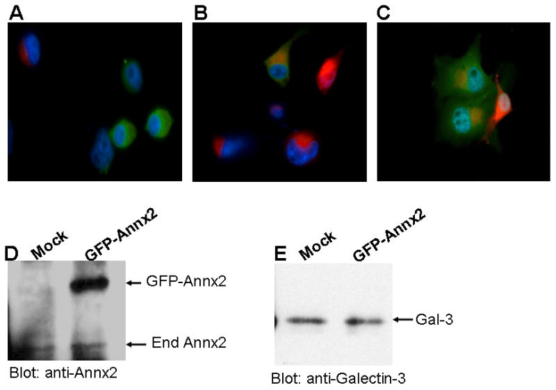
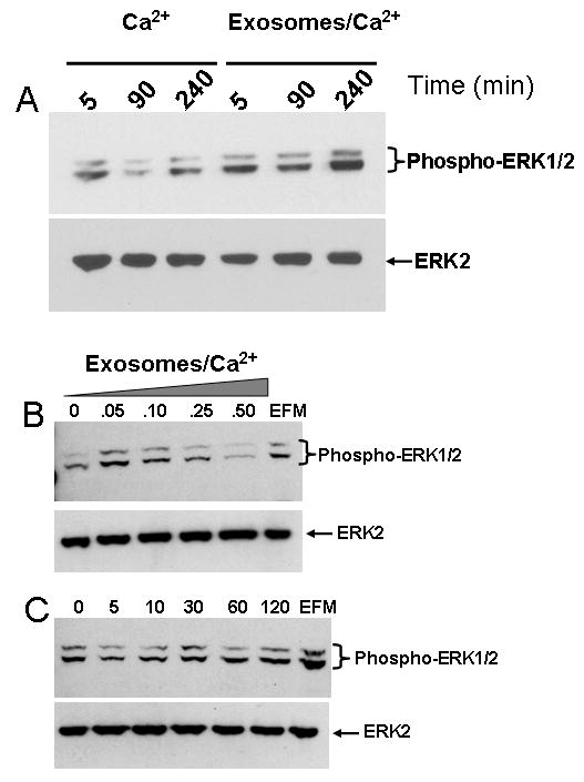
We hereby report studies that suggest a role for serum exosomes in the anchorage-independent growth (AIG) of tumor cells. In AIG assays, fetal bovine serum is one of the critical ingredients. We therefore purified exosomes from fetal bovine serum and examined their potential to promote growth of breast carcinoma cells in soft agar and Matrigel after reconstituting them into growth medium (EEM). In all the assays, viable colonies were formed only in the presence of exosomes. Some of the exosomal proteins we identified, have been documented by others and could be considered exosomal markers. Labeled purified exosomes were up-taken by the tumor cells, a process that could be competed out with excess unlabeled vesicles. Our data also suggested that once endocytosed by a cell, the exosomes could be recycled back to the conditioned medium from where they can be up-taken by other cells. We also demonstrated that low concentrations of exosomes activate MAP kinases, suggesting a mechanism by which they maintain the growth of the tumor cells in soft agar. Taken together, our data demonstrate that serum exosomes form a growth promoting platform for AIG of tumor cells and may open a new vista into cancer cell growth in vivo.


Figures

Figure 1
Figure 1. Bovine serum exosome mediated anchorage independent growth of breast cancer cells in soft agar
Soft agar growth assays were set up in 96-well micro titer plates as described in Materials and Methods. The cells were allowed to grow in soft agar either in the presence (EEM) or absence (EFM) of purified bovine exosomes. After 3 weeks the colonies were either photographed (panel A) or Alamar blue was added to the control wells and to the wells containing exosomes. After 4 hours of color change, the plates were read at 570 nm (panel B). The growth promoting properties of the exosomes were also determined using other breast cancer cell lines (panel C).
Figure 2
Figure 2. Bovine serum exosome mediated growth of stellate colonies of breast cancer cells in Matrigel
Liquid Matrigel (100μl/well) was allowed to solidify at 37°C in 96-well micro titer plate. The carcinoma cells were then added on the Matrigel (500 cells/well) in the presence of serum free medium (panel A), exosome free medium (panel B) and exosome enriched medium (panel C). The cells were then incubated in a humidified CO2 incubator for 14 days and then photographed.
Figure 3
Figure 3. Bovine serum exosome mediated growth of normal and breast cancer cells on plastic
In panel A, MCF-10A were at 2×10⁴ cells/well in 96-well micro titer plates in serum free medium, exosomal enriched medium (EEM) and exosomal free medium (EFM). The cells were allowed to grow for 5 days and then photographed. In panel B, one breast carcinoma cell line, MDA-MB-231 and a melanoma cell line, MDA-MB-435 were likewise plated at 2 × 10⁴ cells/well in 96-well micro titer plates in EEM and EFM and in complete medium (CM) containing 10% fetal bovine serum.
Figure 4
Figure 4. Proteins Associated with bovine serum exosomes
Panel A, transmission electron micrograph of exosomes purified from fetal bovine serum. The black line represents 100 nm. Panel B, aliquots of bovine serum, exosome free serum, and exosomes were resolved in gradient SDS-PAGE and stained with coomassie brilliant blue. The protein bands were photographed and then all the proteins in each lane cut and analyzed by mass spec as described in Materials and Methods. Panel C, proteins from bovine serum, exosome-free serum and exosomes were resolved in SDS-PAGE and then transferred to immobilon paper and probed for fetuin-A and alpha 2 macroglobulin.
Figure 5
Figure 5. Up-take of purified bovine serum exosomes by tumor cells
Purified bovine exosomes were labeled with rhodamine isothiocynate, purified again by ultra-filtration to remove excess unreacted rhodamine and then added to BT-549Gal-3 cells on cover-slips which had been serum starved for 48 h. The cells were incubated with labeled exosomes for 30 min in the absence and presence of excess unlabeled exosomes (0.05 mg/ml) and (0.5 mg/ml) respectively in HBSS containing divalent ions. The cells were rinsed twice with cold PBS and then fixed with -20°C cold methanol for 10 min, cover-slipped and visualized by fluorescent microscopy. The experiment was repeated with MDA-MB-231 and MCF-10A cells with similar results.
Figure 6
Figure 6. Recycling of bovine serum exosomes by tumor cells
BT-549Gal-3 cells were transfected with GFP-annexin-A2 or mock transfected using Fugene6 and serum starved for 48 h. The mock transfected cells were then incubated with rhodamine-labeled exosomes for 30 min at 37°C followed by washing with PBS at room temperature. The GFP-annexin-A2 transfected cells (green cells) and rhodamine labeled (red) cells were harvested with 2 mM EDTA, washed in cold PBS and mixed at a ratio of 1:3 (green cells to red cells), plated in six-well plates on coverslips and incubated for 10 min (Panel A) and then for 2hr at 37°C (Panels B and C). The GFP-annexin-A2 and mock transfected cells were also detached with EDTA and incubated separately in HBSS in siliconized tubes for 2 h at 37°C. Exosomes were isolated and purified from the conditioned medium as described in Materials and Methods. The cellular exosomes were resolved in SDS-PAGE, transferred to immobilon membranes and probed with annexin-2 (Panel D) and galectin-3 (Panel E) antibodies.
Figure 7
Figure 7. MAP Kinase signaling mediated by bovine serum exosomes
BT-549Gal3 cells were plated in 10 cm. dishes and serum starved for 48 h. The adherent cells were incubated in HBS containing 1 mM of the divalent ions (Mg2+/Ca2+) for the indicated time points (Panel A). The cells were scrapped, washed in ice-cold PBS and stored at -80°C until needed. In parallel experiments, cells (2 × 10⁶ cells/dish) were plated on poly(HEMA)-treated dishes in HBS containing the divalent ions (Ca2+/Mg2+) without or with indicated concentrations of exosomes (mg/ml) for 10 min (Panel B). EFM (0.5 mg/ml) was also included as a positive control. In Panel C, the cells were incubated with 0.1 mg/ml of exosomes on poly(HEMA) coated wells for the indicated time points. The EFM (0.1 mg/ml) was also incubated with the cells for 120 min as a control. MAP kinase activation was visualized by detection of phosphorylated ERK1/2 by western blotting using antibodies to the dually phosphorylated ERK1 and ERK2. Total ERK (loading control) was detected using antibodies to ERK2.

References

    1. Klein EA, Yung Y, Castagnino P, Kothapalli D, Assoian RK. Cell adhesion, cellular tension, and cell cycle control. Methods Enzymol. 2007;426:155–175. - PubMed
    1. Schwartz MA. Integrins, oncogenes, and anchorage independence. J Cell Biol. 1997;139:575–578. - PMC - PubMed
    1. Reichelt J. Mechanotransduction of keratinocytes in culture and in the epidermis. Eur J Cell Biol. 2007;86:807–816. - PubMed
    1. Shekhar MP, Pauley R, Heppner G. Host microenvironment in breast cancer development: extracellular matrix-stromal cell contribution to neoplastic phenotype of epithelial cells in the breast. Breast Cancer Res. 2003;5:130–135. - PMC - PubMed
    1. Freedman VH, Shin SI. Cellular tumorigenicity in nude mice: correlation with cell growth in semi-solid medium. Cell. 1974;3:355–359. - PubMed

Publication types