. 2009 Apr;84(4):483-92.
doi: 10.1016/j.ajhg.2009.03.001. Epub 2009 Mar 26.

Duplications involving a conserved regulatory element downstream of BMP2 are associated with brachydactyly type A2

Affiliations

Duplications involving a conserved regulatory element downstream of BMP2 are associated with brachydactyly type A2

Katarina Dathe et al. Am J Hum Genet. 2009 Apr.

Abstract

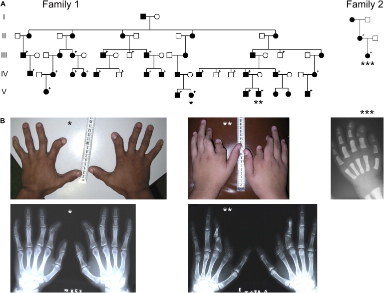
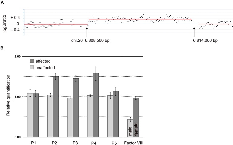
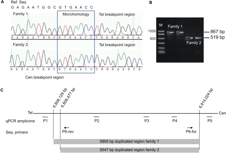
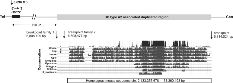
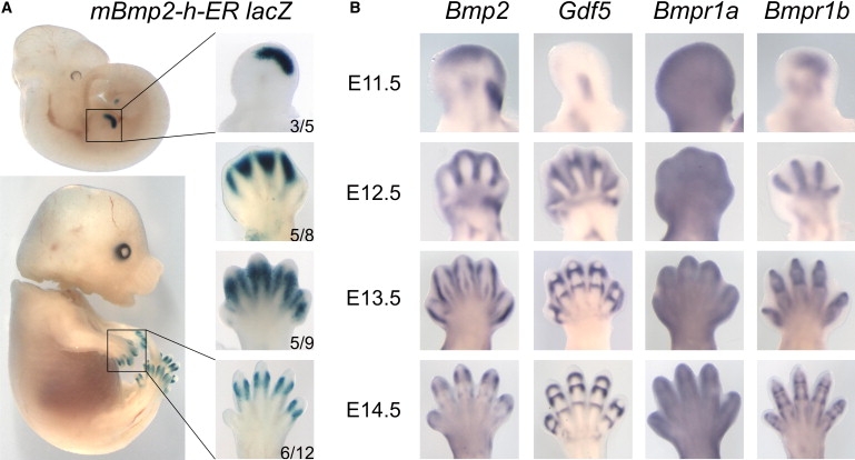
Autosomal-dominant brachydactyly type A2 (BDA2), a limb malformation characterized by hypoplastic middle phalanges of the second and fifth fingers, has been shown to be due to mutations in the Bone morphogenetic protein receptor 1B (BMPR1B) or in its ligand Growth and differentiation factor 5 (GDF5). A linkage analysis performed in a mutation-negative family identified a novel locus for BDA2 on chromosome 20p12.3 that incorporates the gene for Bone morphogenetic protein 2 (BMP2). No point mutation was identified in BMP2, so a high-density array CGH analysis covering the critical interval of approximately 1.3 Mb was performed. A microduplication of approximately 5.5 kb in a noncoding sequence approximately 110 kb downstream of BMP2 was detected. Screening of other patients by qPCR revealed a similar duplication in a second family. The duplicated region contains evolutionary highly conserved sequences suggestive of a long-range regulator. By using a transgenic mouse model we can show that this sequence is able to drive expression of a X-Gal reporter construct in the limbs. The almost complete overlap with endogenous Bmp2 expression indicates that a limb-specific enhancer of Bmp2 is located within the identified duplication. Our results reveal an additional functional mechanism for the pathogenesis of BDA2, which is duplication of a regulatory element that affects the expression of BMP2 in the developing limb.


Figures

Figure 1
Figure 1
Pedigrees and Clinical Phenotype (A) Pedigrees. Affected individuals are indicated by black symbols. Symbols with a + indicate individuals who were clinically examined and for whom further molecular analysis was performed. (B) Clinical phenotype caused by the duplication. BDA2 with shortened and medially deviated second fingers caused by hypoplastic and triangular middle phalanges was observed in almost all of the affected individuals as demonstrated in the photograph and X-rays of an affected adult () and an 18-month-old affected child (∗∗∗). One affected individual in family 1 (∗∗) showed a more severe phenotype, displaying considerable shortened and deviated second and third fingers resulting from hypoplastic and malformed proximal as well as middle phalanges showing similarities to BDC.
Figure 2
Figure 2
Microduplication on 20p12.3 (A) Genomic profile of the microduplication as detected on NimbleGen custom array. The detected breakpoints are indicated by arrows. The duplication comprises ∼5.5 kb. The gray horizontal lines indicate the segments as calculated by the CBS algorithm with 50 bp segmentation. x axis shows genomic positions on chromosome 20; y axis shows log2ratio. (B) Microduplication confirmed by quantitative real-time PCR. The mean values for relative quantification were exported from the 7900 SDS software. For 4 affected (dark gray bars) and 4 nonaffected (light gray bars) individuals, mean values and standard deviations (error bars) for each target amplicon relative to Albumin as a two-copy reference gene were calculated. For gender determination, mean values and SDs for factor VIII on the X chromosome were calculated relative to Albumin as an autosomal two-copy reference gene. Results were calibrated to the mean value determined for a healthy female control. P1–P5 refers to primers shown in Table S1. One duplicated allele plus one normal allele results in three copies for the amplicons of primer pairs P2, P3, and P4 within the duplicated region in the affected individuals and therefore in a ratio of 1.5 relative to the two copies of the healthy female control. Localization of qPCR amplicons is illustrated in Figure 3C.
Figure 3
Figure 3
Breakpoint Identification by Sequence Analysis (A) Sequence analysis of the junction fragment revealed a tandem duplication of 5,895 bp in family 1 and a tandem duplication of 5,547 bp in family 2. The top line on the left displays the centromeric reference sequence (ref. seq.). Below are the electropherograms of one affected individual of each family. The centromeric breakpoint is identical in both families. Note the differences in the telomeric sequence after the seven homologous nucleotides (surrounded by the blue box) of the junction fragment between the two families. The breakpoints are located within this homologous sequence or at the adjacent nucleotides. (B) PCR products obtained with primers P6-forward and P6-reverse result in an 867 bp fragment in family 1 and in a 519 bp fragment in family 2. (C) Illustration of qPCR amplicons and sequencing primer positions as well as localization of the duplicated regions and breakpoints in family 1 and 2 on chromosome 20.
Figure 4
Figure 4
Schematic Representation of the Critical Region on 20p12.3 BMP2 and the 3′ distant duplicated region containing a limb-specific regulatory element that lies about 110 kb downstream of the gene itself are shown. The duplicated region (gray box) in family 1 described here starts at nucleotide 6,808,129 bp and ends at nucleotide 6,814,024 bp on chromosome 20. In family 2, the duplication starts at nucleotide 6,808,477 bp and ends similar as in family 1 at nucleotide 6,808,024 bp (positions according to UCSC Human Genome March 2006). The conservation of the duplicated sequence in different species is depicted in the UCSC genome browser plot. Within the sequence here are two highly conserved regions between mammals and chicken. The positions of the homologous mouse sequence on chromosome 2 are shown under the alignments ranging from 133,355,678 to 133,360,193 bp (UCSC Mouse Genome February 2006). This sequence was cloned into the mBmp2-h-ER_pSfi-Hsp promoter X-Gal transgene construct.
Figure 5
Figure 5
Expression Profiles of X-Gal in Transgenic Mice Compared to Bmp2, Gdf5, and Their Type I Receptors (A) X-Gal staining of transgenic mice carrying the BDA2-associated duplicated region (mBmp2-h-ER). Left panel shows the entire embryo, right panel magnification of fore limb. Developmental stage is given on right side. The number of embryos with specific X-gal staining in the limbs out of the total number of transgene-positive embryos is shown in the right lower corner. Note that staining is present exclusively in the limb autopod. At E11.5, X-Gal staining is present in a distinct region of the distal autopod. Coincidental with the formation of the digit anlagen (E12.5), the staining moves toward the interdigital space. At E13.5 and E14.5, staining becomes restricted to the distal interphalangeal joint region. (B) Whole-mount in situ hybridization of Bmp2, Gdf5, and their receptors Bmpr1a and Bmpr1b. Note overlapping but not identical staining pattern between Bmp2 and X-Gal. Gdf5 expression marks the developing joints. Bmpr1a is expressed ubiquitously throughout the limb, whereas Bmpr1b is restricted to the digit anlagen and the interphalangeal joints.

Comment in

References

    1. Howard M.L., Davidson E.H. cis-Regulatory control circuits in development. Dev. Biol. 2004;271:109–118. - PubMed
    1. Kleinjan D.A., van Heyningen V. Long-range control of gene expression: emerging mechanisms and disruption in disease. Am. J. Hum. Genet. 2005;76:8–32. - PMC - PubMed
    1. Kishigami S., Mishina Y. BMP signaling and early embryonic patterning. Cytokine Growth Factor Rev. 2005;16:265–278. - PubMed
    1. Seemann P., Mundlos S., Lehmann K. Alterations of bone morphogenetic protein signaling pathway(s) in skeletal diseases. In: Vukicevic S., Sampath K.T., editors. Bone Morphogenetic Proteins: From Local to Systemic Therapeutics. Birkhäuser Verlag AG; Basel: 2008. pp. 141–159.
    1. Lehmann K., Seemann P., Stricker S., Sammar M., Meyer B., Suring K., Majewski F., Tinschert S., Grzeschik K.H., Muller D. Mutations in bone morphogenetic protein receptor 1B cause brachydactyly type A2. Proc. Natl. Acad. Sci. USA. 2003;100:12277–12282. - PMC - PubMed

Publication types

MeSH terms