Review
. 2009 Jun 5;583(11):1728-35.
doi: 10.1016/j.febslet.2009.03.049. Epub 2009 Mar 27.

Glycoproteomics: past, present and future

Affiliations
Review

Glycoproteomics: past, present and future

Bérangère Tissot et al. FEBS Lett. .

Abstract

This invited paper reviews the study of protein glycosylation, commonly known as glycoproteomics, beginning with the origins of the subject area in the early 1970s shortly after mass spectrometry was first applied to protein sequencing. We go on to describe current analytical approaches to glycoproteomic analyses, with exemplar projects presented in the form of the complex story of human glycodelin and the characterisation of blood group H eptitopes on the O-glycans of gp273 from Unio elongatulus. Finally, we present an update on the latest progress in the field of automated and semi-automated interpretation and annotation of these data in the form of GlycoWorkBench, a powerful informatics tool that provides valuable assistance in unravelling the complexities of glycoproteomic studies.


Figures

Figure 1
Figure 1
A simplified glycoproteomic experimental workflow is shown, illustrating common approaches to glycoproteomic analysis. Samples take the form of slices or spots excised from single or multi-dimensional polyacrylamide gels, or batches of cells, fluids, immunoprecipitates or tissue extracts. Analytical approaches can broadly be categorised as “top down” or “bottom up”. The former, illustrated by a purple arrow, begins with work on purified samples of glycoproteins in an attempt to identify the intact molecular weight profile by direct MALDI-TOF MS or by ES-MS. By subtracting the known or inferred mass of the protein component, the type and extent of glycosylation may then be deduced. In “bottom up” approaches, which incidentally are essential for describing the detailed glycosylation profile of any protein, the glycoprotein is digested enzymatically and/or chemically, ideally with high-specificity procedures, and the resulting peptide/glycopeptide mixture is mapped mass spectrometrically either by on-line (red arrows) LC-ES-MS followed by MS/MS analysis of signals of interest, or by off-line (blue arrows) strategies involving ES- or MALDI-MS and MS/MS approaches. Prior to these separation and mapping procedures, various strategies for enrichment of glycopeptides may be introduced, including lectin binding (see text). Parallel glycomic analyses (illustrated by the green arrow) are an invaluable feature of the “bottom up” approach involving enzymatic or chemical release of the glycans followed by MALDI-TOF MS or ES-MS mapping of the glycan populations, usually as permethyl derivatives (see text), providing information about specific glycans and their relative amounts, which can then be compared and matched with data at the glycopeptide and overall glycoprotein levels.
Figure 2
Figure 2
Nano-LC-ES-MS of a tryptic digest of a human glycodelin sample. The sample was reduced, carboxymethylated and dialysed prior to digestion with trypsin. A few micrograms of the digest products were subjected to on-line reverse-phase nanoLC coupled to a Q-STAR mass spectrometer. Panel A shows the total ion chromatogram (TIC) for a 90-min experiment. Extracted ion chromatograms of glycan fragment ions were used to determine when glycopeptides elute and Panel B shows the summation of all spectra acquired between 7.5 and 11 min which were identified as corresponding to a glycopeptide elution time window. The diagnostic glycan fragment ions are annotated in the low-mass region of the spectrum shown in Panel B. Panels C, D and E detail portions of the spectrum with special emphasis at middle mass (Panel D) and high mass (Panel E) which have been expanded to show typical patterns for mixtures of multiply charged glycopeptides. For simplicity, the majority of the signals in the complete spectrum are not labelled but all multiply charged signals can be attributed to glycoforms of two major peptides containing the same N-glycosylation site at Asn-63. Sugar symbols used throughout this chapter are those employed by the Consortium for Functional Glycomics. Circles represent hexoses (yellow: Galactose, green: Mannose), squares represent N-acetylhexosamines (yellow: N-acetylgalactosamine, blue: N-acetylglucosamine), red triangle: Fucose, purple diamond: N-acetylneuraminic acid.
Figure 3
Figure 3
MALDI-TOF MS profiling of Unio elongatulus glycoprotein gp273 O-glycans. Around 250 micrograms of gp273 were reduced, carboxymethylated and digested with trypsin. N-glycans were removed using PNGase A then separated from the pool of peptides and O-glycopeptides. The latter were subjected to reductive elimination and reduced O-glycans were purified prior to being permethylated and analysed using MALDI-TOF instrumentation. The Unio elongatulus genome is unknown so far. Therefore information about possible glycan structures cannot be readily deduced from biosynthetic knowledge. The use of a semi-automated tool such as GlycoWorkbench proved to be essential for an unbiased and comprehensive annotation of this MS spectrum. The putative compositions shown in boxes on this figure have been obtained using the GlycoPeakFinder feature of GlycoWorkbench (see Text). The type of monosaccharides considered were restricted to Hexose (10 maximum, symbol: blank circle), N-acetylhexosamine (10 maximum, symbol: blank square), Pentose (1 maximum, symbol: blank star), deoxy-Hexose (10 maximum, symbol: blank triangle), Hexuronic Acid (3 maximum, symbol: blank 2-halved-diamond) and Neuraminic acid (1 Maximum, symbol: blank diamond).
Figure 4
Figure 4
GlycoWorkbench annotated MALDI-TOF/TOF MS/MS spectrum of the Unio elongatulus O-glycan species at m/z 912 (panel A) and 1680 (panel B) obtained using MALDI TOF/TOF instrumentation. According to the semi-automated annotation performed using GycoPeakFinder the species at m/z 912 and 1680 could correspond to 2 and 6 compositions, respectively. For each of the two MS/MS spectra, a list of fragment ions was manually selected. This list was then compared with the lists of predicted ions calculated for each of the possible structures corresponding to the computed compositions. A report showing the scores for each proposed arrangement was produced and the highest score glycan structures are shown on the top right corner of the spectra. Once the structures are selected, GlycoWorkbench can generate annotated spectra as shown here. For monosaccharide keys, see Figure 2. The reduced reducing end is represented by the formula image 
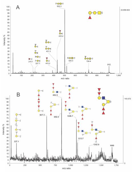
symbol. Keys related to fragmentation are the following: formula image represents a fragmentation on the reducing end side of the glycosidic bond (also known as B or Z ions) and formula image represents a fragmentation on the non-reducing end side of the glycosidic bond (also known as C or Y ions).

References

    1. Dell A, Morris HR. Glycoprotein structure determination by mass spectrometry. Science. 2001;291:2351–6. - PubMed
    1. Morris HR, Chalabi S, Panico M, Sutton-Smith M, Clark GF, Goldberg D, Dell A. Glycoproteomics: Past, present and future. International Journal of Mass Spectrometry. 2007;259:16–31.
    1. Morris HR, Thompson MR, Osuga DT, Ahmed AI, Chan SM, Vandenheede JR, Feeney RE. Antifreeze glycoproteins from the blood of an antarctic fish. The structure of the proline-containing glycopeptides. J Biol Chem. 1978;253:5155–62. - PubMed
    1. Taylor GW, Morris HR, Peterson TE, Magnusson S. Advances in Mass Spectrometry. 1980;18:1090.
    1. Morris HR, Panico M, Taylor GW. FAB-mapping of recombinant-DNA protein products. Biochem Biophys Res Commun. 1983;117:299–305. - PubMed

Publication types