. 2009 May;15(5):958-67.
doi: 10.1261/rna.669209. Epub 2009 Mar 27.

A conformational switch in the DiGIR1 ribozyme involved in release and folding of the downstream I-DirI mRNA

Affiliations

A conformational switch in the DiGIR1 ribozyme involved in release and folding of the downstream I-DirI mRNA

Henrik Nielsen et al. RNA. 2009 May.

Abstract

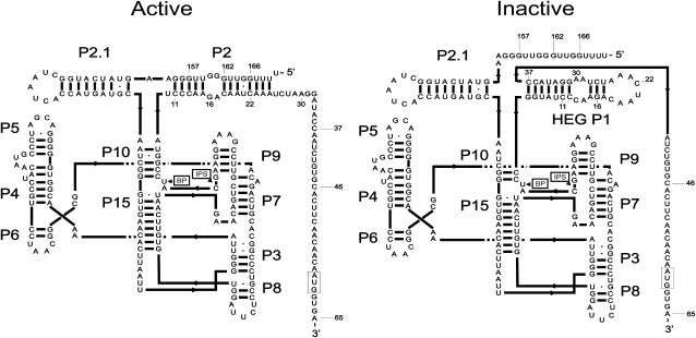
DiGIR1 is a group I-like cleavage ribozyme found as a structural domain within a nuclear twin-ribozyme group I intron. DiGIR1 catalyzes cleavage by branching at an Internal Processing Site (IPS) leading to formation of a lariat cap at the 5'-end of the 3'-cleavage product. The 3'-cleavage product is subsequently processed into an mRNA encoding a homing endonuclease. By analysis of combinations of 5'- and 3'-deletions, we identify a hairpin in the 5'-UTR of the mRNA (HEG P1) that is formed by conformational switching following cleavage. The formation of HEG P1 inhibits the reversal of the branching reaction, thus giving it directionality. Furthermore, the release of the mRNA is a consequence of branching rather than hydrolytic cleavage. A model is put forward that explains the release of the I-DirI mRNA with a lariat cap and a structured 5'-UTR as a direct consequence of the DiGIR1 branching reaction. The role of HEG P1 in GIR1 branching is reminiscent of that of hairpin P-1 in splicing of the Tetrahymena rRNA group I intron and illustrates a general principle in RNA-directed RNA processing.


Figures

FIGURE 1.
FIGURE 1.
Helix diagrams of the catalytically active conformation of DiGIR1 compared with an alternative, inactive conformation. The difference between the two is the engagement of P10″ and P2″ (the 3′-strands of the two helices) of the catalytically active conformation in formation of HEG P1 in the inactive conformation. HEG P1 is also found as a structural element in the 5′-UTR of the I-DirI mRNA. It is possible that the inactive conformation has additional structural differences from the active conformation than HEG P1 emphasized in the figure. The branch point (BP) and internal processing site (IPS) are indicated, and the initiation codon of the HE open reading frame is boxed. The numbers indicate the positions upstream or downstream, respectively, in relation to the IPS. These positions mark the number of nucleotides included in the deletion constructs analyzed in the paper.
FIGURE 2.
FIGURE 2.
(A) Reactions that are considered in the interpretation of the cleavage analyses: (1A) branching reaction in which the precursor is cleaved at IPS, resulting in a 5′-product with a 3′-OH and a 3′-product with a 5′-lariat cap in which the first and the third nucleotides are joined by a 2′, 5′-phosphodiester bond (Nielsen et al. 2005); (1B) reversal of the branching reaction; (2A) cleavage at the IPS by hydrolysis resulting in a 5′-product with a 3′-OH and a 3′-product with a 5′-phosphate; (2B) reverse of the hydrolytic cleavage reaction; (3A) hydrolytic opening of the lariat; (3B) formation of the lariat directly from the 5′-cleavage product. The thickness of the arrows indicates the efficiency of the isolated reactions. (Dashed arrows) Reactions that have not been observed in the present experiments. (B) Influence of HEG P1 on ligation. Ligation of unlabeled 166-nt 5′-cleavage fragment with labeled 3′-cleavage fragment of variable lengths. Time samples were withdrawn at t = 0, 2, 15, and 60 min. (C). Effect of mutation of HEG P1 on ligation. The experiment was conducted similarly to the experiment described above except that an acidic ligation buffer was used in order to inhibit the forward reaction and that more time samples were withdrawn for analysis (t = 0, 1, 2, 5, 10, 30, 60, and 120 min). The effect of the mutations on the base pairing potential in HEG P1 is shown below the gel picture.
FIGURE 3.
FIGURE 3.
DiGIR1 cleavage and primer extension analyses of length variants of DiGIR1 with 166 nt (A), 162 nt (B), and 157 nt (C) included upstream of the IPS. (Graphs) Cleavage analyses; (autoradiograms) primer extension analyses. Numbers refer to the positions indicated in Fig. 1. The figure in B was constructed using data from Einvik et al. (2000).
FIGURE 4.
FIGURE 4.
Analysis of the effect of urea on a standard DiGIR1 cleavage reaction. (A) Gel analysis (6% UPAG) of cleavage of DiGIR1-162.22 without and with the addition of urea to a final concentration of 2 M. (Pre) Precursor transcript. The 3′-product of the cleavage reaction is not included in the figure. (B) Quantitative analysis of the autoradiogram shown in A. (Inset) Calculated k obs and end-point values. (C) Primer extension analysis of aliquots of the same time samples analyzed in A. (BP) Branch Point, (IPS) Internal Processing Site. A sequencing ladder made by sequencing of a DiGIR1 plasmid with the same primer as used for primer extension is shown to the left. (D). Effect of urea on the reversal of the branching reaction (ligation). Gel-purified 5′- and 3′-cleavage fragments were used for the experiment. The 5′-fragment was unlabeled and the 3′-fragment radioactively labeled during transcription. Urea was added to a concentration of 2 M and samples were withdrawn at t = 0, 10, and 30 min.
FIGURE 5.
FIGURE 5.
Accessibility of structural elements P2, P2.1, and P10 to kethoxal modification prior to and after cleavage. (A) Primer extension analysis of modified RNA prior to cleavage (Pre [162.65]) and after cleavage by hydrolysis (Post [166.65]) or branching (Post [162.65]). A sequencing ladder made by sequencing of a DiGIR1 plasmid with the same primer as used for primer extension is shown to the left. (B) Helix diagrams showing the structure of the P2-P2.1-P10 domain prior to (Pre) and after (Post) cleavage. The nucleotides involved in forming HEG P1 are italicized. The observed modifications are indicated in the post-cleavage structure. None of these structures (indicated by arrows) were found to be modified in the precursor RNA (Pre).
FIGURE 6.
FIGURE 6.
Cleavage analysis of three different 5′-length variants analyzed in parallel on native (A) and 6% denaturing (urea) polyacrylamide (B) gels. The cleavage reactions were incubated for 0, 5, 30, and 120 min, respectively. The 5′- and 3′-products are indicated.
FIGURE 7.
FIGURE 7.
(A) Comparison of features of conformational change between the Tetrahymena group I splicing ribozyme and the Didymium GIR1 branching ribozyme. (B) Schematic outline of the conformational switch involved in I-DirI mRNA maturation. In the precursor transcript (top), HEG P1 is formed, resulting in an inactive DiGIR1 ribozyme. After DiGIR2 catalyzed self-splicing, a conformational switch reorganizes the HEG P1 residues into the active form of DiGIR1 with subsequent cleavage by branching at the internal processing site (IPS). HEG P1 then reappears in the mature I-DirI mRNA (bottom) along with a lariat cap and a poly(A) tail at the 5′- and 3′-ends, respectively. (SSU) Small subunit ribosomal RNA exon, (5′- and 3′-SS) 5′- and 3′-splice sites, (G) exogenous guanosine cofactor added at the 5′-end of the excised intron RNA during self-splicing.

Similar articles

Cited by

References

    1. Agback P., Sandstrom A., Yamakage S., Sund C., Glemarec C., Chattopadhyaya J. Solution structure of lariat RNA by 500 MHz NMR spectroscopy and molecular dynamics studies in water. J. Biochem. Biophys. Methods. 1993;27:229–259. - PubMed
    1. Badorrek C.S., Gherghe C.M., Weeks K.M. Structure of an RNA switch that enforces stringent retroviral genomic RNA dimerization. Proc. Natl. Acad. Sci. 2006;103:13640–13645. - PMC - PubMed
    1. Beckert B., Nielsen H., Einvik C., Johansen S.D., Westhof E., Masquida B. Molecular modelling of the GIR1 branching ribozyme gives new insight into evolution of structurally related ribozymes. EMBO J. 2008;27:667–678. - PMC - PubMed
    1. Cao Y., Woodson S.A. Destabilizing effect of an rRNA stem–loop on an attenuator hairpin in the 5′ exon of the Tetrahymena pre-rRNA. RNA. 1998;4:901–914. - PMC - PubMed
    1. Cao Y., Woodson S.A. Refolding of rRNA exons enhances dissociation of the Tetrahymena intron. RNA. 2000;6:1248–1256. - PMC - PubMed

Publication types

LinkOut - more resources