. 2009;4(3):e5061.
doi: 10.1371/journal.pone.0005061. Epub 2009 Mar 30.

A central role for Foxp3+ regulatory T cells in K-Ras-driven lung tumorigenesis

Affiliations

A central role for Foxp3+ regulatory T cells in K-Ras-driven lung tumorigenesis

Courtney A Granville et al. PLoS One. 2009.

Abstract

Background: K-Ras mutations are characteristic of human lung adenocarcinomas and occur almost exclusively in smokers. In preclinical models, K-Ras mutations are necessary for tobacco carcinogen-driven lung tumorigenesis and are sufficient to cause lung adenocarcinomas in transgenic mice. Because these mutations confer resistance to commonly used cytotoxic chemotherapies and targeted agents, effective therapies that target K-Ras are needed. Inhibitors of mTOR such as rapamycin can prevent K-Ras-driven lung tumorigenesis and alter the proportion of cytotoxic and Foxp3+ regulatory T cells, suggesting that lung-associated T cells might be important for tumorigenesis.

Methods: Lung tumorigenesis was studied in three murine models that depend on mutant K-Ras; a tobacco carcinogen-driven model, a syngeneic inoculation model, and a transgenic model. Splenic and lung-associated T cells were studied using flow cytometry and immunohistochemistry. Foxp3+ cells were depleted using rapamycin, an antibody, or genetic ablation.

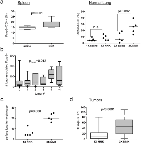
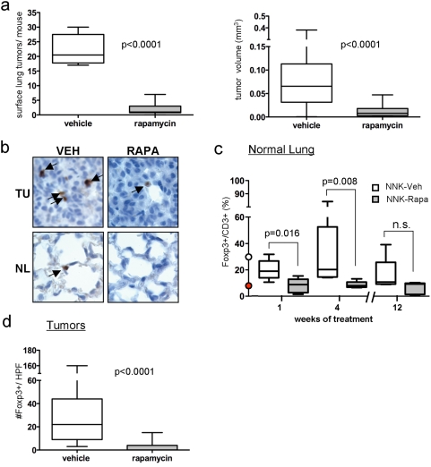
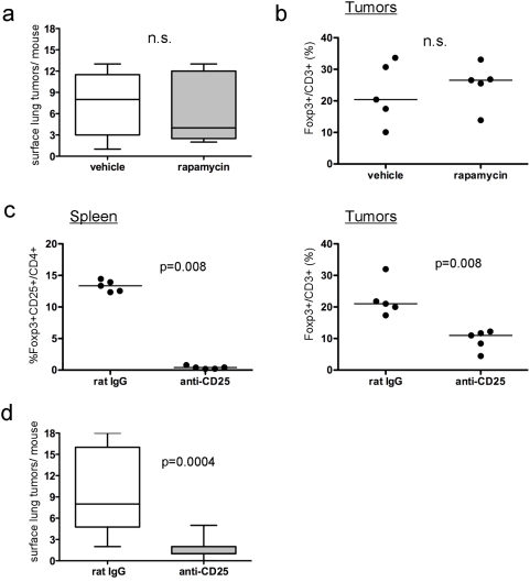
Results: Exposure of A/J mice to a tobacco carcinogen tripled lung-associated Foxp3+ cells prior to tumor development. At clinically relevant concentrations, rapamycin prevented this induction and reduced lung tumors by 90%. In A/J mice inoculated with lung adenocarcinoma cells resistant to rapamycin, antibody-mediated depletion of Foxp3+ cells reduced lung tumorigenesis by 80%. Likewise, mutant K-Ras transgenic mice lacking Foxp3+ cells developed 75% fewer lung tumors than littermates with Foxp3+ cells.

Conclusions: Foxp3+ regulatory T cells are required for K-Ras-mediated lung tumorigenesis in mice. These studies support clinical testing of rapamycin or other agents that target Treg in K-Ras driven human lung cancer.


Conflict of interest statement

Competing Interests: The authors have declared that no competing interests exist.

Figures

Figure 1
Figure 1. NNK increases Foxp3+ cells.
(a) Assessment of Foxp3+ cells in splenic and lung tissues. One week after NNK exposure, Foxp3+ T cell subsets were assessed in spleens (left) as a fraction of total CD4+ splenocytes and in lungs (right) as a fraction of total CD3+ cells. (b) Correlation of number of NNK-induced lung tumors with number of Foxp3+ cells in surrounding lung tissues. Tumors were counted in mice 16 wk after NNK administration, and scoring for Foxp3+ cells was performed using IHC. A trend for the number of Foxp3+ cells in surrounding normal lung with the number of tumors per lung is shown. (c–d) Dose-dependent induction of lung tumors (c) and Foxp3+ cells (d) by NNK. For (a, b, and d), boxes indicate interquartile range, lines indicate median, and whiskers indicate minimal and maximal values. For (a and c), each point represents a mouse and the line represents the median. A high-powered field (HPF) indicates a field under 100× magnification.
Figure 2
Figure 2. Rapamycin prevents NNK-induced tumorigenesis and depletes lung-associated Foxp3+ cells in NNK-treated mice.
(a) Tumor multiplicity (left) and tumor size (right) after 12 wk of rapamycin or vehicle. (b) Representative staining for Foxp3+ cells in tumors (TU) and normal airway epithelium (NL). (c) The fraction of Foxp3+/total CD3+ cells in normal lung tissues from saline or NNK-exposed mice treated with rapamycin or vehicle for 1, 4 or 12 weeks was calculated after IHC was performed. The red and white dots at week 0 indicate the percentages of Foxp3+/CD3+ cells in the lungs of A/J mice prior to and after NNK administration, respectively. (d) The number of Foxp3+ cells in tumors arising 16 weeks after NNK exposure in the presence or absence of rapamycin was determined using IHC. For (a, c, and d), boxes indicate interquartile range, lines indicate median, and whiskers indicate minimal and maximal values. A high-powered field (HPF) indicates a field under 100× magnification.
Figure 3
Figure 3. Depletion of Foxp3+ cells using an anti-CD25 antibody decreases the ability of rapamycin-resistant lung adenocarcinoma cells to form tumors.
(a–b) Effect of rapamycin on IO33 tumor multiplicity (a) or tumor-associated percent of Foxp3+/CD3+ cells (b). (c) Effect of rat IgG or anti-CD25 antibodies on the number of splenic Foxp3+CD25+/CD4+ cells (left) and IO33 tumor-associated %Foxp3+/CD3+ cells (right). (d) Effect of anti-CD25 antibody or rat IgG on IO33 lung tumor multiplicity. For (a) and (d), boxes indicate interquartile range, lines indicate median, and whiskers indicate minimal and maximal values. For (b–c), each point represents a mouse and the line represents the median. n.s., not significant.
Figure 4
Figure 4. Genetically engineered mice that lack Foxp3+ cells develop fewer K-Ras driven lung tumors.
(a) Representative IHC of tumor-associated Foxp3+ and CD3+ cells in tobacco-carcinogen (NNK) and K-rasLA2 transgenic mouse models of lung tumorigenesis. (b) Quantification of IHC analysis in (a) for tumor-associated percent of Foxp3+/CD3+ cells. Each point represents a mouse and the line represents the median; n.s., not significant. (c) Tumor multiplicity in K-RASLA2/wt/Foxp3/Y or K-RASLA2/wt/Foxp3+/Y offspring. Boxes indicate interquartile range, lines indicate median, and whiskers indicate minimal and maximal values.

References

    1. Jemal A, Siegel R, Ward E, Hao Y, Xu J, et al. Cancer statistics, 2008. CA Cancer J Clin. 2008;58:71–96. - PubMed
    1. IARC. Tobacco smoking and tobacco smoke. IARC Monographs on the Evaluation of the Carcinogenic Risks of Chemicals to Humans. Lyon, France: International Agency for Research on Cancer; 2004. - PMC - PubMed
    1. Westra WH, Slebos RJ, Offerhaus GJ, Goodman SN, Evers SG, et al. K-ras oncogene activation in lung adenocarcinomas from former smokers. Evidence that K-ras mutations are an early and irreversible event in the development of adenocarcinoma of the lung. Cancer. 1993;72:432–438. - PubMed
    1. Westra WH, Baas IO, Hruban RH, Askin FB, Wilson K, et al. K-ras oncogene activation in atypical alveolar hyperplasias of the human lung. Cancer Res. 1996;56:2224–2228. - PubMed
    1. Mills NE, Fishman CL, Rom WN, Dubin N, Jacobson DR. Increased prevalence of K-ras oncogene mutations in lung adenocarcinoma. Cancer Res. 1995;55:1444–1447. - PubMed

Publication types