

Automated simulation of areal bone mineral density assessment in the distal radius from high-resolution peripheral quantitative computed tomography
- PMID: 19330422
- PMCID: PMC2777210
- DOI: 10.1007/s00198-009-0907-0
Automated simulation of areal bone mineral density assessment in the distal radius from high-resolution peripheral quantitative computed tomography
Abstract
Summary: An automated image processing method is presented for simulating areal bone mineral density measures using high-resolution peripheral quantitative computed tomography (HR-pQCT) in the ultra-distal radius. The accuracy of the method is validated against clinical dual X-ray absorptiometry (DXA). This technique represents a useful reference to gauge the utility of novel 3D quantification methods applied to HR-pQCT in multi-center clinical studies and potentially negates the need for separate forearm DXA measurements.
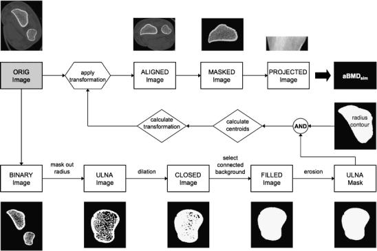
Introduction: Osteoporotic status is primarily assessed by measuring areal bone mineral density (aBMD) using 2D dual X-ray absorptiometry (DXA). However, this technique does not sufficiently explain bone strength and fracture risk. High-resolution peripheral quantitative computed tomography (HR-pQCT) has been introduced as a method to quantify 3D bone microstructure and biomechanics. In this study, an automated method is proposed to simulate aBMD measures from HR-pQCT distal radius images.
Methods: A total of 117 subject scans were retrospectively analyzed from two clinical bone quality studies. The distal radius was imaged by HR-pQCT and DXA on one of two devices (Hologic or Lunar). Areal BMD was calculated by simulation from HR-pQCT images (aBMD(sim)) and by standard DXA analysis (aBMD(dxa)).
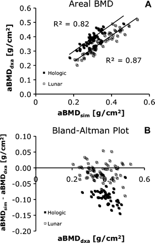
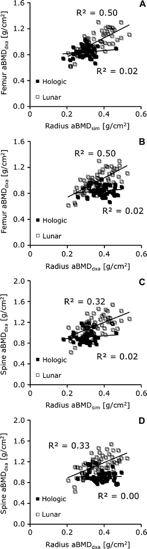
Results: The reproducibility of the simulation technique was 1.1% (root mean-squared coefficient of variation). HR-pQCT-based aBMD(sim) correlated strongly to aBMD(dxa) (Hologic: R (2) = 0.82, Lunar: R (2) = 0.87), though aBMD(sim) underestimated aBMD(dxa) for both DXA devices (p < 0.0001). Finally, aBMD(sim) predicted aBMD at the proximal femur and lumbar spine with equal power compared to aBMD(dxa).
Conclusion: The results demonstrate that aBMD can be simulated from HR-pQCT images of the distal radius. This approach has the potential to serve as a surrogate forearm aBMD measure for clinical HR-pQCT studies when axial bone mineral density values are not required.
Figures
References
-
- (1991) Consensus development conference: prophylaxis and treatment of osteoporosis. Am J Med 90:107-110. - PubMed
-
- Black DM, Thompson DE. The effect of alendronate therapy on osteoporotic fracture in the vertebral fracture arm of the Fracture Intervention Trial. Int J Clin Pract Suppl. 1999;101:46–50. - PubMed
Publication types
MeSH terms
Grants and funding
LinkOut - more resources
Full Text Sources
Other Literature Sources
Medical
