. 2009 Dec;5(4):461-71.
doi: 10.1007/s11302-009-9148-0. Epub 2009 Mar 28.

ATP as a mediator of macula densa cell signalling

Affiliations

ATP as a mediator of macula densa cell signalling

P Darwin Bell et al. Purinergic Signal. 2009 Dec.

Abstract

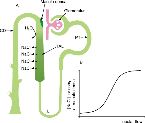
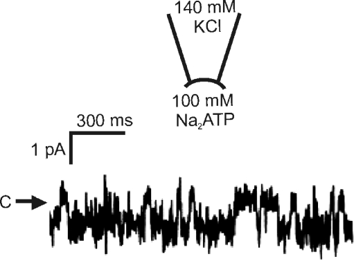
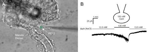
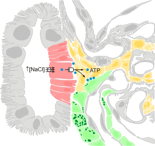
Within each nephro-vascular unit, the tubule returns to the vicinity of its own glomerulus. At this site, there are specialised tubular cells, the macula densa cells, which sense changes in tubular fluid composition and transmit information to the glomerular arterioles resulting in alterations in glomerular filtration rate and blood flow. Work over the last few years has characterised the mechanisms that lead to the detection of changes in luminal sodium chloride and osmolality by the macula densa cells. These cells are true "sensor cells" since intracellular ion concentrations and membrane potential reflect the level of luminal sodium chloride concentration. An unresolved question has been the nature of the signalling molecule(s) released by the macula densa cells. Currently, there is evidence that macula densa cells produce nitric oxide via neuronal nitric oxide synthase (nNOS) and prostaglandin E(2) (PGE(2)) through cyclooxygenase 2 (COX 2)-microsomal prostaglandin E synthase (mPGES). However, both of these signalling molecules play a role in modulating or regulating the macula-tubuloglomerular feedback system. Direct macula densa signalling appears to involve the release of ATP across the basolateral membrane through a maxi-anion channel in response to an increase in luminal sodium chloride concentration. ATP that is released by macula densa cells may directly activate P2 receptors on adjacent mesangial cells and afferent arteriolar smooth muscle cells, or the ATP may be converted to adenosine. However, the critical step in signalling would appear to be the regulated release of ATP across the basolateral membrane of macula densa cells.


Figures

Fig. 1
Fig. 1
a Schematic representation of a nephro-vascular unit. The major nephron segments are identified; PT proximal tubule, LH loop of Henle, TAL thick ascending limb, and CD collecting duct. b Relationship between flow rate through the nephron and the sodium chloride concentration and osmolality of the tubular fluid at the macula densa
Fig. 2
Fig. 2
A multiphoton confocal image of an isolated perfused JGA preparation in which both afferent arteriole and thick ascending limb are perfused. Note the close anatomical association between the macula densa plaque and the afferent arteriole. Also, the green fluorescence is quinacrine, which stains acidic vesicles and most likely represents renin-containing granules
Fig. 3
Fig. 3
Wide-field image of an isolated perfused thick ascending limb. The glomerulus is not shown because fura-2, the calcium-sensitive fluorophore, was loaded through the lumen. High levels of cytosolic calcium are red and yellow, while lower values are green and the lowest calcium concentrations are in blue. The macula densa cells have a much lower cytosolic calcium concentration compared to the surrounding epithelial cells
Fig. 4
Fig. 4
Representative trace recorded from an inside-out patch at the basolateral membrane of a macula densa cell. C and arrow indicate the closed current level. Current is mediated by a maxi-anion channel and the downward deflections reflect movement of ATP through the channel
Fig. 5
Fig. 5
a Photomicrograph showing the isolated perfused thick ascending limb-glomerular preparation. The glomerulus has been partially removed to allow access to the basolateral membrane. A biosensor (in this case a PC12 cell) is then placed at the basolateral membrane of these cells. ATP-P2X activity in the biosensor cell is measured with fura-2 or using whole-cell patch-clamp. Biosensor cell is pseudo-coloured green for identification. (b) Whole-cell patch-clamp of the biosensor cell showing the effects of increasing luminal sodium chloride concentration; these results are consistent with the activation of a P2X channel
Fig. 6
Fig. 6
Schematic representation of the macula densa cell signalling pathway in which ATP release by the macula densa cells plays a critical and essential role. Macula densa cells, extra/intraglomerular mesangial cells and vascular smooth muscle cells of the afferent arteriole are shown in red, yellow and green, respectively

References

    1. Barajas L (1979) Anatomy of the juxtaglomerular apparatus. Am J Physiol 237:F333–F343 - PubMed
    1. Schafer JA, Troutman SL, Andreoli TE (1974) Volume reabsorption, transepithelial potential differences, and ionic permeability properties in mammalian superficial proximal straight tubules. J Gen Physiol 64:582–607 - PMC - PubMed
    1. Burg MB, Green N (1973) Function of the thick ascending limb of Henle's loop. Am J Physiol 224:659–668 - PubMed
    1. Rocha AS, Kudo LH (1982) Water, urea, sodium, chloride, and potassium transport in the in vitro isolated perfused papillary collecting duct. Kidney Int 22:485–491 - PubMed
    1. Barajas L, Salido EC, Liu L, Powers KV (1995) The juxtaglomerular apparatus: a morphologic perspective. In: Laragh JH et al (eds) Hypertension: pathophysiology, diagnosis and management. Raven, New York, pp 1335–1348