. 2009 Apr 15;315(7):1234-46.
doi: 10.1016/j.yexcr.2009.01.021. Epub 2009 Feb 3.

Live-cell imaging demonstrates extracellular matrix degradation in association with active cathepsin B in caveolae of endothelial cells during tube formation

Affiliations

Live-cell imaging demonstrates extracellular matrix degradation in association with active cathepsin B in caveolae of endothelial cells during tube formation

Dora Cavallo-Medved et al. Exp Cell Res. .

Abstract

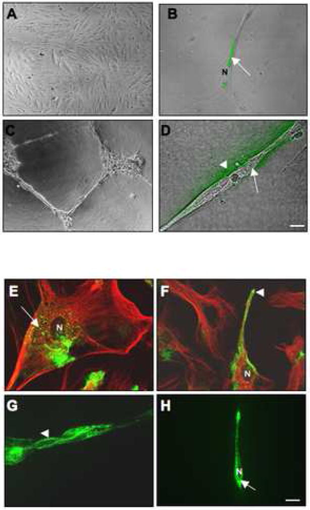
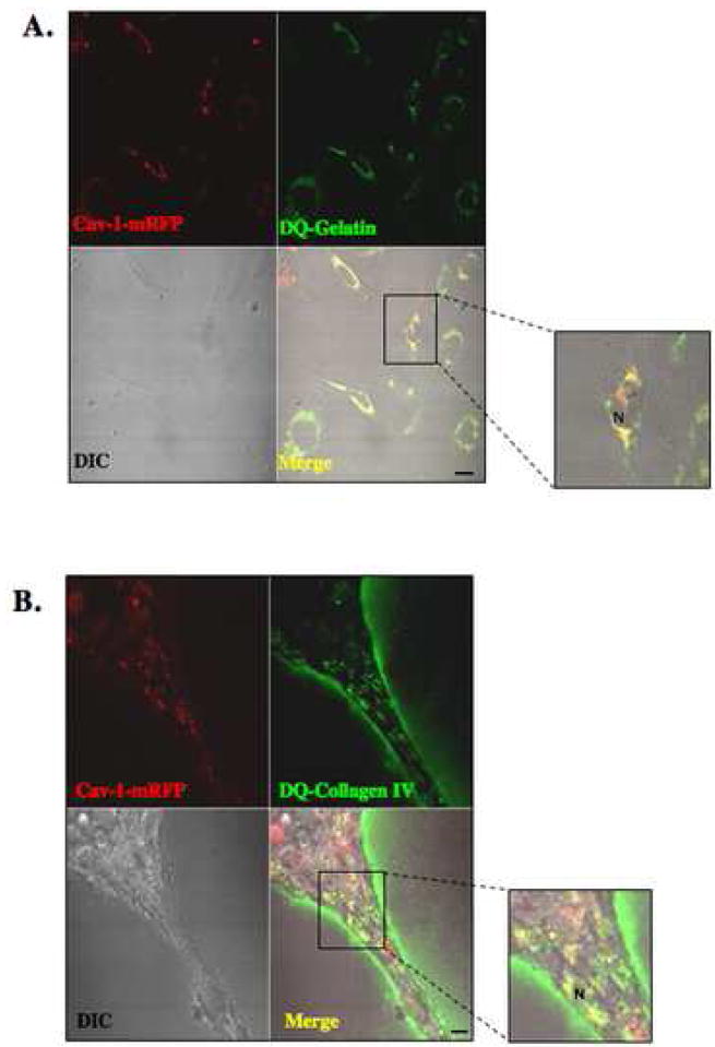
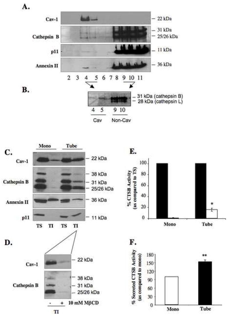
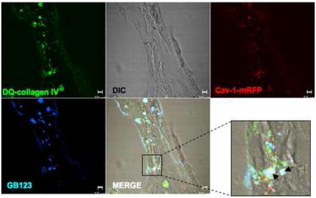
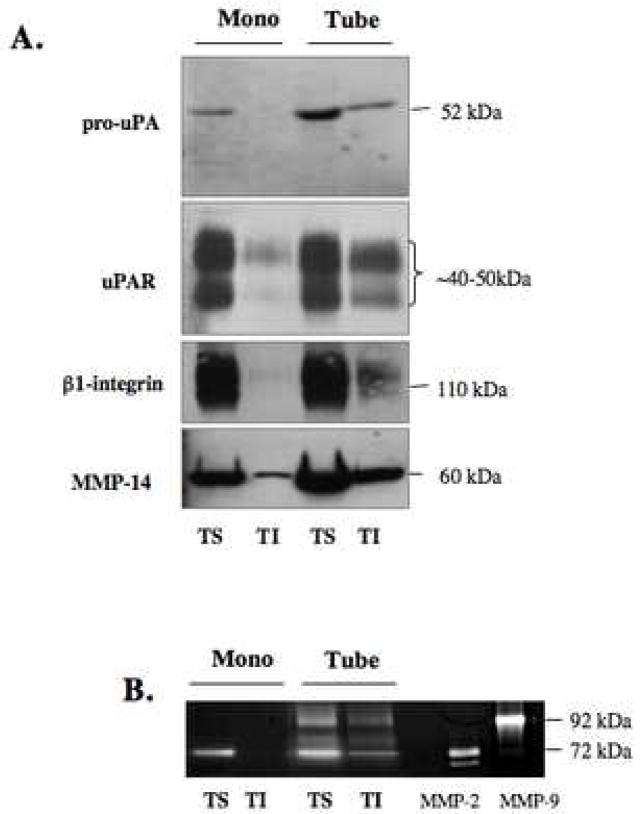
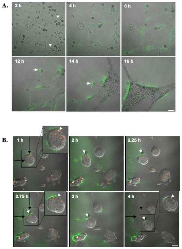
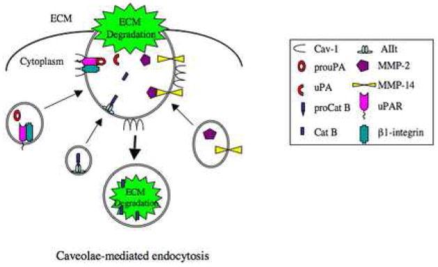
Localization of proteases to the surface of endothelial cells and remodeling of the extracellular matrix (ECM) are essential to endothelial cell tube formation and angiogenesis. Here, we partially localized active cathepsin B and its cell surface binding partners, S100A/p11 (p11) of the annexin II heterotetramer (AIIt), to caveolae of human umbilical vein endothelial cells (HUVEC). Via a live-cell proteolysis assay, we observed that degradation products of quenched-fluorescent (DQ)-proteins (i.e. gelatin and collagen IV) colocalized intracellularly with caveolin-1 (cav-1) of HUVEC grown in either monolayer cultures or in vitro tube formation assays. Activity-based probes that bind covalently to active cysteine cathepsins and degradation products of DQ-collagen IV partially localized to intracellular vesicles that contained cav-1 and active cysteine cathepsins. Biochemical analyses revealed that the distribution of active cathepsin B in caveolar fractions increased during in vitro tube formation. Pro-uPA, uPAR, MMP-2 and MMP-14, which have been linked with cathepsin B to ECM degradation pathways, were also found to increase in caveolar fractions during in vitro tube formation. Our findings are the first to demonstrate through live-cell imaging ECM degradation in association with active cathepsin B in caveolae of endothelial cells during tube formation.


Figures

Figure 1
Figure 1. Gelatin and collagen IV degradation by and localization of endogenous cav-1 in HUVEC grown as a monolayer versus tube-like structures
Equal numbers of HUVEC were grown for 16 h on glass coverslips coated with gelatin alone (A), gelatin containing 25 μg/ml DQ-gelatin (B), rBM (Cultrex) alone (C), or rBM containing 25 μg/ml DQ-collagen IV (D). After 16 h, confocal images were taken of live cells [differential interference contrast (DIC)] and DQ-substrate degradation products (green), which are present intracellularly (arrow) in HUVEC grown on gelatin (B) and both intracellularly (arrow) and pericellularly (arrowhead) in HUVEC grown on rBM (D). Immunostaining for endogenous cav-1 using rabbit anti-human caveolin antibodies was performed on fixed and permeabilized HUVEC cells grown on gelatin (E and F; two different fields of view) or rBM (G and H; two different fields of view). The morphology of HUVEC in monolayer culture is apparent from the immunostaining for tubulin (red) using mouse anti-human tubulin antibodies (E and F). Staining for caveolin is observed in the perinuclear region (arrow) and at the cell surface (arrowhead). N, nucleus. Bar, 20 μm.
Figure 2
Figure 2. Intracellular colocalization of cav-1 with gelatin and collagen IV degradation products in HUVEC
Equal numbers of HUVEC transfected with cav-1-mRFP construct were grown for 16 h on glass coverslips coated with either gelatin containing 25 μg/ml DQ-gelatin (A) or rBM (Cultrex) containing 25 μg/ml DQ-collagen IV (B). After 16 h, confocal images were taken of live cells (DIC), cav-1-mRFP (red) and DQ-substrate degradation products (green). DQ-substrate degradation products (green) are present intracellularly in HUVEC grown on gelatin and both intracellularly and pericellularly in HUVEC grown on rBM (see inserts). Colocalization of cav-1-mRFP and DQ-substrate degradation products appears yellow in the merged images. N, nucleus. Bar, 20 μm.
Figure 3
Figure 3. Increased distribution of active cathepsin B and p11 to caveolae of HUVEC cells during tube formation
HUVEC grown as a monolayer were subjected to subcellular fractionation on a sucrose gradient following homogenization in sodium carbonate buffer, pH 11.0. One ml fractions were collected from the top of the gradient and equal volume aliquots of fractions 2–11 were analyzed by SDS-PAGE and immunoblotted with anti-caveolin, anti-cathepsin B, anti-annexin II and anti-p11 antibodies (A). Aliquots from caveolae fractions 4 and 5 (Cav) and non-caveolae fractions 9 and 10 (non-Cav) isolated from HUVEC treated with 1μM GB111-FL were analyzed by SDS-PAGE and imaged using a Typhoon laser scanner. Cathepsin B (31 kDa) and cathepsin L (28 kDa) were labeled (B). Equal numbers of HUVEC were grown on gelatin (mono) or rBM (tube) for 18 h and caveolae were isolated by successive detergent extraction as described in Materials and Methods. Triton-soluble (TS) represents non-caveolae fractions and Triton-insoluble (TI) represents caveolae fractions. Equal volume aliquots of each fraction were analyzed by SDS-PAGE and immunoblotted with anti-caveolin, anti-cathepsin B, anti-annexin II and anti-p11 antibodies (C). To verify distribution of caveolae to TI fractions, HUVEC grown for 18 h on rBM were treated with 10 mM MβCD for 1 h at 37 °C prior to caveolae isolation (D). TS (closed bars) and TI (open bars) fractions of HUVEC monolayers (mono) and tube-like structures (tube) were assayed for cathepsin B (CTSB) activity against Z-Arg-Arg-NHMec substrate and activity was recorded as pmol/min/cell number. The specific activity for caveolae-associated cathepsin B activity is expressed as a percentage of cathepsin B activity in the non-caveolar fraction (TS) (E). Overnight conditioned media of HUVEC grown as a monolayer on gelatin (mono) and during tube formation (tube) were also analyzed for cathepsin B activity and expressed as a percentage of secreted activatable cathepsin B activity from monolayer HUVEC (F). Immunoblots and graphs, presented as mean ± standard deviation, are representative of at least three experiments. * P< 0.05 and ** P< 0.01.
Figure 4
Figure 4. Active cathepsin B partially localizes to caveolae of HUVEC during tube formation
HUVEC were transiently transfected with a cav-1-mRFP construct and grown on glass coverslips coated with rBM containing 25 μg/ml DQ-collagen IV in the presence of 1 μM GB-123 for 18 h. Thereafter, unbound GB-123 was washed away and cells incubated 1–2 h in probe-free medium. Confocal images were then taken of live cells (DIC), cav-1-mRFP (red), DQ-collagen IV degradation products (green) and GB-123 bound to cysteine cathepsins (pseudocolored blue). Colocalization of cav-1-mRFP and DQ-collagen IV degradation products appears yellow, colocalization of DQ-collagen IV degradation products and GB-123 appears pale blue, and colocalization of cav-1-mRFP, DQ-collagen IV degradation products and GB-123 appears white (arrows; see insert) in the merged image. Bar, 10 μm.
Figure 5
Figure 5. Increased distribution of pro-uPA, uPAR, β1-integrin, MMP-2 and MMP-14 to HUVEC caveolae during tube formation
Equal numbers of HUVEC were grown on gelatin (mono) or rBM (tubes) for 18 h and caveolae were isolated by successive detergent extraction as described in Materials and Methods. Triton-soluble (TS) represents non-caveolae fractions and Triton-insoluble (TI) represents caveolae fractions. Equal volume aliquots of each fraction were analyzed by SDS-PAGE and immunoblotted with anti-uPA, anti-uPAR, anti-β1-integrin and anti-MMP-14 antibodies (A). Equal volume aliquots of each fraction were also analyzed by gelatin zymography for MMP-2 and MMP-9 activities (D). Purified MMP-2 (10 ng) and MMP-9 (5 ng) were used as positive controls. Immunoblots and zymograms are representative of at least three experiments.
Figure 6
Figure 6. Time courses for collagen IV proteolysis and colocalization of collagen IV degradation products with cav-1 during HUVEC tube formation
HUVEC were grown on glass coverslips coated with rBM containing 25 μg/ml DQ-collagen IV. (A) Confocal images were taken of live cells between 2 and 16 h. DQ-collagen IV degradation products (green) are seen surrounding tubular structures and at the rear of a migrating endothelial cell (arrow). Cell sprouting is observed at 2h (arrowheads). Bar, 100 μm. (B) Confocal images were taken between 1 and 4 h of live HUVEC transfected with cav-1-mRFP. DQ-collagen IV degradation products (green) and cav-1 (red) are seen colocalized (yellow) intracellularly (arrowheads) and pericellular degradation of DQ-collagen IV is observed at the rear of a migrating endothelial cell (arrows). Black arrows represent the original location of a migrating HUVEC cell (in box) at 1h. Bar, 20 μm.
Figure 7
Figure 7. Potential protease network in endothelial cell caveolae during ECM degradation
In endothelial cells, proteases (i.e., cathepsin B, pro-uPA and MMPs) and their associated proteins/receptors (i.e., AIIt, uPAR and β1-integrin) are translocated to cell surface caveolae via intracellular vesicles (i.e., multivesicular bodies and exosomes) [14]. Caveolae-associated proteases are activated and degrade ECM proteins both pericellularly and intracellularly via caveolae-mediated endocytosis.

References

    1. Risau W. Mechanisms of angiogenesis. Nature. 1997;386:671–674. - PubMed
    1. van Hinsbergh VW, Engelse MA, Quax PH. Pericellular proteases in angiogenesis and vasculogenesis. Arterioscler Thromb Vasc Biol. 2006;26:716–728. - PubMed
    1. Bergers G, Brekken R, McMahon G, Vu TH, Itoh T, Tamaki K, Tanzawa K, Thorpe P, Itohara S, Werb Z, Hanahan D. Matrix metalloproteinase-9 triggers the angiogenic switch during carcinogenesis. Nat Cell Biol. 2000;2:737–744. - PMC - PubMed
    1. Pepper MS. Role of the matrix metalloproteinase and plasminogen activator-plasmin systems in angiogenesis. Arterioscler Thromb Vasc Biol. 2001;21:1104–1117. - PubMed
    1. Rakic JM, Maillard C, Jost M, Bajou K, Masson V, Devy L, Lambert V, Foidart JM, Noel A. Role of plasminogen activator-plasmin system in tumor angiogenesis. Cell Mol Life Sci. 2003;60:463–473. - PMC - PubMed

Publication types