. 2009 Jun;13(3):519-29.
doi: 10.1016/j.media.2009.02.006. Epub 2009 Mar 5.

Vessel target location estimation during the TIPS procedure

Affiliations

Vessel target location estimation during the TIPS procedure

Guillaume Piliere et al. Med Image Anal. 2009 Jun.

Abstract

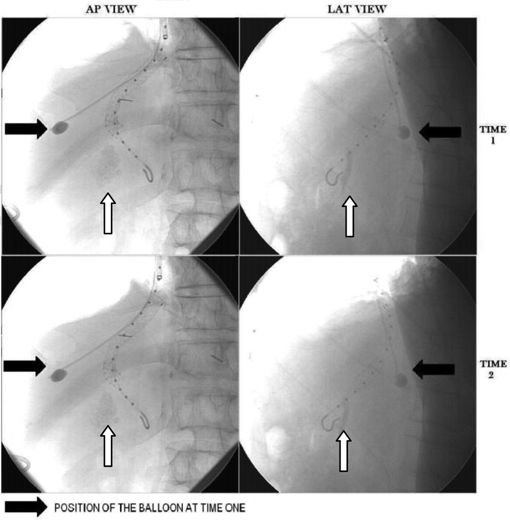
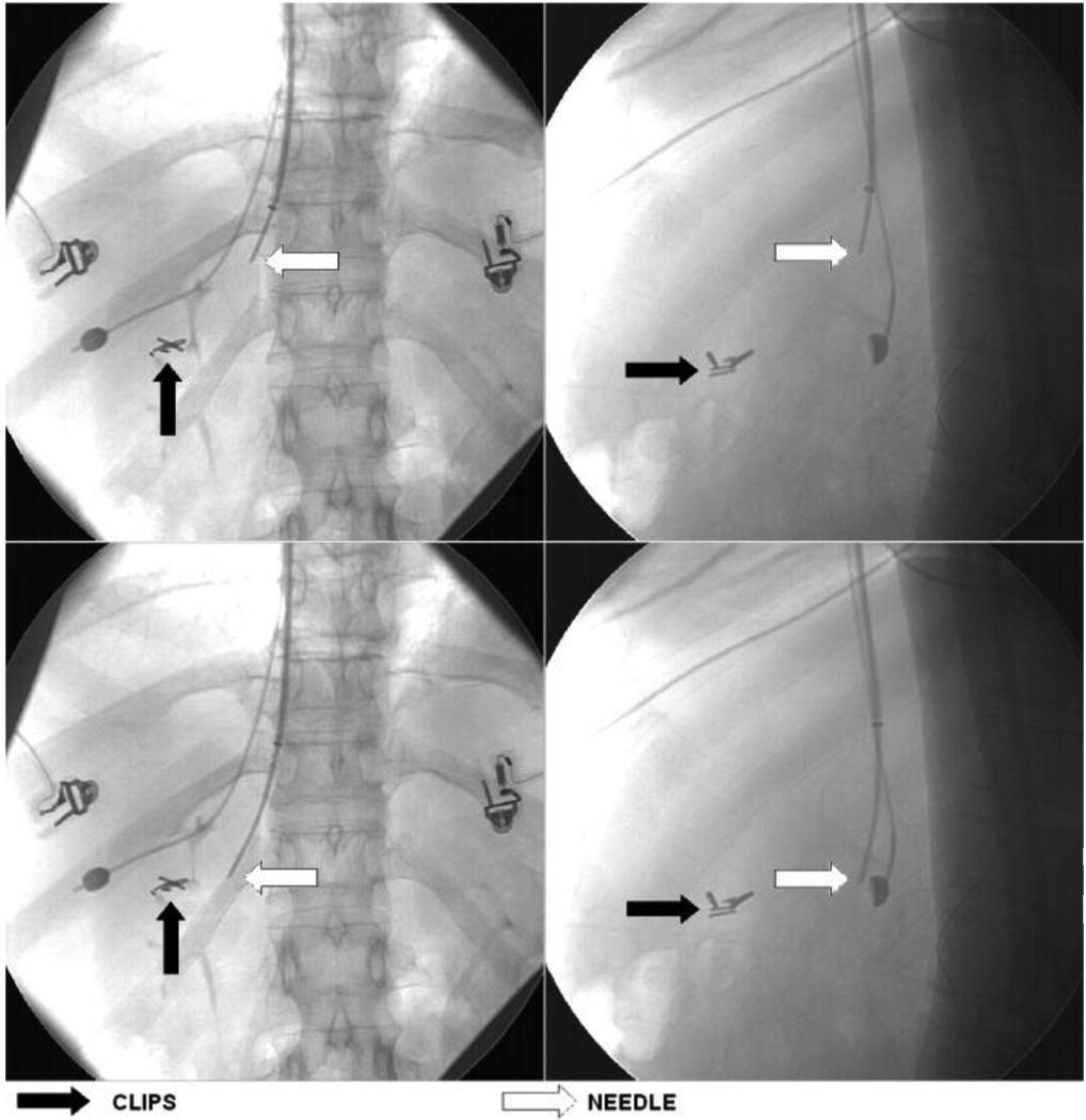
Creation of a transjugular intrahepatic portosystemic shunt (TIPS) requires passage of a needle toward a moving target that is only seen transiently by X-ray prior to needle passage. Intraoperative, 3D target localization would facilitate target access and improve the safety of the procedure. The clinical assumption is that patients undergoing the TIPS procedure possess rigid, cirrhotic livers that undergo only intraoperative translation without significant deformation or rotation. Based upon this assumption, we hypothesize that the position of any unseen, 3D target point within the liver can be determined intraoperatively by precalculation of the relative positions of the target point to a different 3D point that can be tracked intraoperatively. This paper examines this hypothesis using intraoperatively acquired, biplane, X-ray images of seven patients. In six, we tracked the effects of cardiac and respiratory motion, and in three the effects of needle pressure. Methods involved reconstruction of 3D vessel bifurcation and other trackable intrahepatic points from biplane angiograms, measurement of liver deformation by examining changing distances between these 3D points over time, and comparison of expected to actual displacements of these points with respect to a fixed reference point in the liver. We conclude that, for the rigid livers associated with patients undergoing TIPS, that there is less intraoperative deformation than previously reported by other groups addressing healthy liver deformation, and that the location of an unseen target can be predicted within 3mm accuracy.


Figures

Fig. 1
Fig. 1
Overview of the procedure. A: Drawing of the liver, chest and hepatic venous system (blue) and the portal venous system (gold). The goal of the TIPS procedure is to create an artificial connection between the hepatic and portal veins. B. X-ray image with opacification of hepatic vein with injected contrast material (dark) with overlay of a static, preacquired, subtracted image of the portal and hepatic venous systems (white) prior to needle insertion. Thin white arrow points to the portal veins and white arrowhead points to hepatic veins at time 1, and black arrowhead points to hepatic veins at time 2. Note the superior translation of the hepatic veins (black at time 2) due to respiratory motion between the two image acquisitions.
Fig. 2
Fig. 2
Simplified overview of the approach illustrated by a single AP image at time 1 (left) and time 2 (right). Black ball = projection of the opacified balloon. Gold and blue stars = projections of “point blue” and “point gold” Gold arrow = vector between balloon and “point gold”. Blue arrow = vector between balloon and “point blue”. Red arrow = distance between points blue and gold. The opacified balloon (black) has moved downward at time 2. If there is no liver deformation the distances between all three points should remain constant at times 1 and 2 (the lengths of the red, gold, and blue arrows should remain constant). If it is possible to use the opacified balloon to track the new position of “point blue” at time 2, the blue vector (determined at time 1) when added to the new balloon position at time 2 should correctly point to “point blue” at time 2. Similarly, the new position of “point gold” should be predicted by adding the gold vector defined at time 1 to the new balloon position at time 2. Errors induced by liver rotation or deformation can be estimated by comparing the predicted to the actual locations of points blue and gold. This figure illustrates a simplified approach in 2D; our study employed all vector direction and length calculations in 3D.
Figure 3
Figure 3
AP (left) and lateral (right) angiograms obtained at time 1 (top row) and subtracted images obtained at time 2 (bottom row). The solid black arrows point to the balloon in each image. The white arrows point to one of the several identified vessel branchpoints. Note that on the subtracted images at time 2 that both the original balloon position is evident (white disc, black arrow) as well as the new balloon position (dark disc, black arrow). There is obvious displacement of the balloon as well as of the identified branchpoint (white arrow) between times 1 and 2.
Figure 4
Figure 4
AP (left) and lateral (right) views of balloon, catheter within the deployed TIPS stent, and opacified gall bladder obtained at times 1 (top row) and 2 (bottom row). The black arrow shows the position of the balloon at time 1. The white arrow points to the partially opacified gall bladder. The catheter within the TIPS stent is the curved line with multiple dots along its length. There is downward motion of the balloon, gallbladder, and stent between times 1 and 2.
Fig. 5
Fig. 5
AP (left) and Lat (right) images of the needle push study for patient with clips (black arrow), needle (white arrow) and balloon at time 1 (top) and time 2 (bottom).

References

    1. Anand BS. Cirrhosis of liver. World J Med. 1999;171:110–115. - PMC - PubMed
    1. Aylward S, Bullitt E. Initialization, noise, singularities and scale in height ridge traversal for tubular object centerline extraction. IEEE-TMI. 2002;21:61–75. - PubMed
    1. Baert SAM, van de Kraats EB, van Walsum T, Viergever MA. Three-Dimensional Guide-Wire Reconstruction from Biplane Image Sequences for Integrated Display in 3-D Vasculature. IEEE TMI. 2003;22:1252–1258. - PubMed
    1. Banares R, Casado M, Rodriques JM, Camunez F, Matilla A, Echenagusia A, Simo G, Piqueras B, Clemente G, Co E. Urgent transjugular intrahepatic portosystemic shunt for control of acute variceal bleeding. Am J Gastroenter. 1998;93:75–79. - PubMed
    1. Banovac F, Tang J, Xu S, Lindisch D, Chung HY, Levy EB, Chang T, McCullough MF, Yaniv Z, Wood BJ, Cleary K. Precision targeting of liver lesions using a novel electromagnetic navigation device in physiologic phantom and swine. Medical Physics. 2005;32:2698–2705. - PubMed

Publication types

MeSH terms