. 2009 Jul;57(7):675-85.
doi: 10.1369/jhc.2009.953380. Epub 2009 Mar 30.

Assessment of PARP-3 distribution in tissues of cynomolgous monkeys

Affiliations

Assessment of PARP-3 distribution in tissues of cynomolgous monkeys

Michèle Rouleau et al. J Histochem Cytochem. 2009 Jul.

Abstract

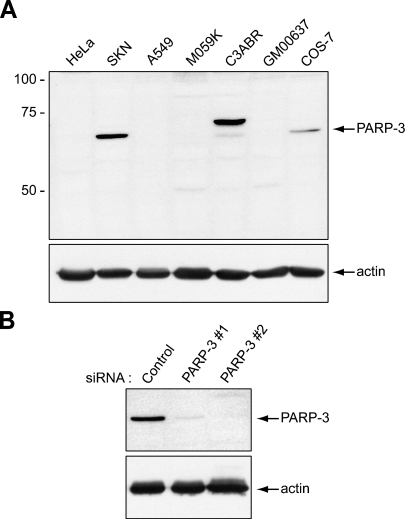
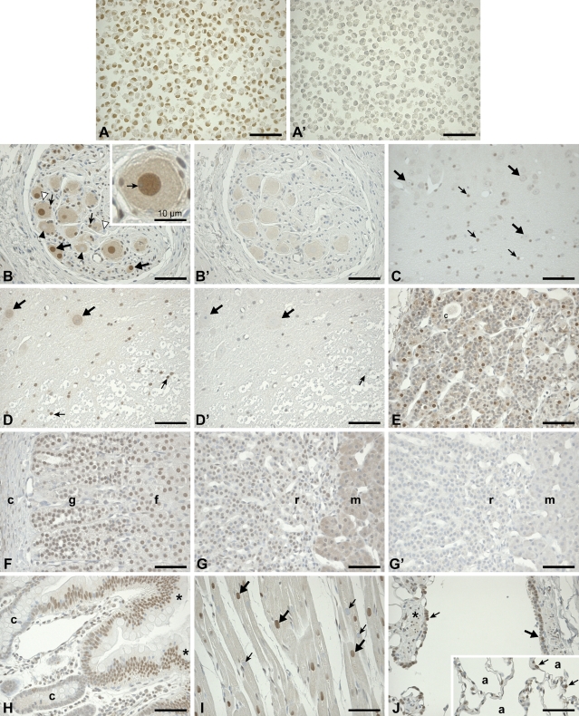
Poly(ADP-ribose) polymerase 3 (PARP-3) is a newly characterized PARP. In contrast to the two best-studied nuclear PARPs, PARP-1 and PARP-2, PARP-3 activity is apparently not stimulated by DNA damage. However, our previous work has demonstrated that PARP-3 interacts with several DNA damage response proteins, including Ku70/Ku80, DNA-PK, and PARP-1, suggesting that it contributes to the DNA damage response. Furthermore, a possible function for PARP-3 in the regulation of gene expression has been inferred from our observations that it associates with polycomb group proteins, which are responsible for epigenetic modifications leading to gene silencing. In this report, we extend our characterization of PARP-3 by revealing its distribution in the tissues and cell types of adult cynomolgous monkeys using a well-characterized PARP-3 polyclonal antibody. This study is the first to demonstrate that PARP-3 is genuinely expressed in most of the examined tissues. However, its expression is highly restricted to specific cell types of each tissue, indicating that PARP-3 expression is tightly regulated. One of the key findings of this study is that PARP-3 is highly expressed in the nuclei of epithelial cells forming the ducts of prostate, salivary glands, liver, and pancreas and in the neurons of terminal ganglia.


Figures

Figure 1
Figure 1
Validation of the specificity of the poly(ADP-ribose) polymerase 3 (PARP-3) antibody by Western blot analysis. (A) Top panel: The anti–PARP-3 antibody detects a single protein band of 62 kDa in the human cell line SK-N-SH and in the monkey cell line COS-7. In the C3ABR human cell line, both PARP-3 isoforms, of 67 kDa and 62 kDa, are detected (see text). The other cell lines do not express detectable levels of PARP-3. Lower panel: A duplicate blot was probed with an anti-actin antibody to show equal loading. (B) Top panel: In SK-N-SH cells transfected with small interfering RNAs (siRNAs) targeting PARP-3 (PARP-3#1 or PARP-3#2), levels of the 62-kDa protein detected with the PARP-3 antibody are drastically reduced. Levels of the 62-kDa protein are not affected by transfection with a control siRNA. Lower panel: A duplicate blot was probed with an anti-actin antibody to show equal loading.
Figure 2
Figure 2
Expression of PARP-3 in normal monkey tissues. (A) Validation of the specificity of the PARP-3 antibody in paraffin section of the human cell line SK-N-SH. The nuclei of all cells are strongly labeled with the anti–PARP-3 antibody. (A′) Preincubation of the PARP-3 antibody with an excess of the immunogenic peptide abolishes staining of the consecutive SK-N-SH paraffin section, demonstrating the specificity of the antibody. (B) A strong nuclear staining is detected in many neurons of terminal ganglia (thick arrows) and satellite cells (thin arrows). However, a number of adjacent neurons and satellite cells are not labeled (black and white arrowheads, respectively). Nucleoli are not labeled, as indicated by the arrow in the inset. (B′) In the control adjacent section, no labeling is detected in the nuclei. (C) In the brain stem, several nuclei of neuroglial cells are labeled (thin arrows), whereas the neurons are not labeled (thick arrows). (D) In the gray matter of spinal cord, nuclei of some neurons are weakly labeled (thick arrows), whereas in the white matter, neuroglial cells are moderately labeled (thin arrows). (D′) No labeling is observed in the consecutive section of the spinal cord labeled with the preadsorbed antibody. Arrows indicate the same cells as in (D). (E) In the pituitary gland, a strong nuclear staining is detected in the pars tuberalis. The weak cytoplasmic staining is specific. Colloidal material (c) is seen within a cord of cells. (F) In the adrenal gland, nuclei of zona glomerulosa (g) and fasciculata (f) cells are moderately labeled. Cells of the capsule (c) are not labeled. (G) In cells of the zona reticularis (r), nuclei are weakly labeled. In the medulla (m), the cytoplasm of parenchymal cells is lightly but specifically labeled. (G′) In the adjacent control section, no staining could be detected. (H) In the duodenum, most of the absorptive epithelial cells lining the villi (asterisk) are labeled in the nucleus, whereas the glandular epithelia of the crypts (c) are not labeled. (I) The nuclei of several cardiac muscle cells are strongly labeled (thick arrows), whereas some of the nuclei of their neighbor cells are completely void of the labeling (thin arrows). (J) In the terminal and respiratory bronchioles of the lungs, some of the ciliated cells (thick arrow) and Clara cells (thin arrow) are moderately labeled. Nuclei of smooth-muscle cells (asterisks) are also labeled. In the alveolar walls (inset), nuclei of several septal cells (thin arrows) lining alveoli (a) are moderately labeled. Bar = 30 μM unless otherwise stated.
Figure 3
Figure 3
Expression of PARP-3 in glandular tissues and reproductive organs of male and female monkeys. (A) In the pancreas, several nuclei of cells in the islets of Langerhans are moderately labeled with the anti–PARP-3 (arrows), whereas acinar cells are not labeled (arrowhead). Centroacinar cells (small arrowheads) forming an intercalated duct are labeled. (B) Most nuclei of the pancreatic duct cells are labeled, as shown in one of the large collecting ducts. (C) In the salivary glands, most of the nuclei of intercalated duct cells (thin arrow) as well as the nuclei of the larger striated duct (thick arrow) and excretory duct (asterisk) cells are labeled. Acinar cells (arrowheads) are not labeled. (D) In the liver, nuclei of epithelial cells lining the bile ducts (arrows) are labeled, whereas nuclei of hepatocytes (arrowheads) are devoid of staining. The light staining seen in the cytoplasm of hepatocytes is not specific, because it persisted in the corresponding control section (not shown). (E) In renal corpuscles of the kidney, nuclei of several podocytes (arrows), as well as nuclei of some endothelial cells, are labeled. Most of the nuclei of the epithelial cells lining collecting ducts (CT) and proximal convoluted tubules (pt) are labeled, whereas no immunostaining reaction could be detected in cells of the distal convoluted tubules (dc). (F) In the thymus, nuclei of several epithelial reticular supporting cells (thick arrows) and of Hassall's corpuscle cells (thin arrows) are labeled. Lymphocytes (arrowheads) are not labeled. (G) In the epidermis of the skin, the nuclei of some basal cells are weakly to moderately labeled (arrows), whereas the upper epidermal cell nuclei are less labeled. (H) Many nuclei of sebaceous gland cells (arrowheads) are labeled. (I) In the testis, the nuclei of Leydig cells (thick arrow) and some of the nuclei of blood vessels (thin arrow) of the interstitial tissue are labeled. In the seminiferous tubules (st), none of the spermatogenic cells are labeled (white arrowheads), whereas most of Sertoli cell nuclei are weakly labeled (black arrowheads). (J) In the prostate, the majority of epithelial cell nuclei of the cranial lobe (arrows) are weakly labeled, whereas (K) most nuclei of cells lining the large prostatic ducts (arrows) as well as of many stromal cells (arrowheads) are strongly labeled. (L) In the corpus of the epididymis, most nuclei of ductal epithelial cells are moderately labeled. (M) In the ovary, some nuclei of follicular cells of a primordial follicle are labeled (thick arrow), whereas the nucleus of the oocyte is not labeled (thin arrow). The weak staining of the germinal epithelium (asterisks) is not specific, because labeling persisted in the control section (not shown). (N) The nuclei of granulosa lutein cells of the corpus luteum show strong labeling (thick arrows) but not the nuclei of theca lutein cells (thin arrows). (O) In the villi of the oviduct, most of the nuclei of epithelial lining cells are moderately labeled but not the nuclei of connective tissue cells (ct). Bar = 30 μM.

References

    1. Augustin A, Spenlehauer C, Dumond H, Menissier-De Murcia J, Piel M, Schmit AC, Apiou F, et al. (2003) PARP-3 localizes preferentially to the daughter centriole and interferes with the G1/S cell cycle progression. J Cell Sci 116:1551–1562 - PubMed
    1. Concha II, Figueroa J, Concha MI, Ueda K, Burzio LO (1989) Intracellular distribution of poly(ADP-ribose) synthetase in rat spermatogenic cells. Exp Cell Res 180:353–366 - PubMed
    1. Haince JF, Kozlov S, Dawson VL, Dawson TM, Hendzel MJ, Lavin MF, Poirier GG (2007) Ataxia telangiectasia mutated (ATM) signaling network is modulated by a novel poly(ADP-ribose)-dependent pathway in the early response to DNA-damaging agents. J Biol Chem 282:16441–16453 - PubMed
    1. Haince JF, McDonald D, Rodrigue A, Dery U, Masson JY, Hendzel MJ, Poirier GG (2008) PARP1-dependent kinetics of recruitment of MRE11 and NBS1 proteins to multiple DNA damage sites. J Biol Chem 283:1197–1208 - PubMed
    1. Hassa PO, Hottiger MO (2008) The diverse biological roles of mammalian PARPS, a small but powerful family of poly-ADP-ribose polymerases. Front Biosci 13:3046–3082 - PubMed

Publication types

Substances