Review
. 2009 May 15;587(Pt 10):2151-61.
doi: 10.1113/jphysiol.2009.171595. Epub 2009 Mar 30.

Gating of the CFTR Cl- channel by ATP-driven nucleotide-binding domain dimerisation

Affiliations
Review

Gating of the CFTR Cl- channel by ATP-driven nucleotide-binding domain dimerisation

Tzyh-Chang Hwang et al. J Physiol. .

Abstract

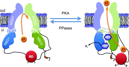
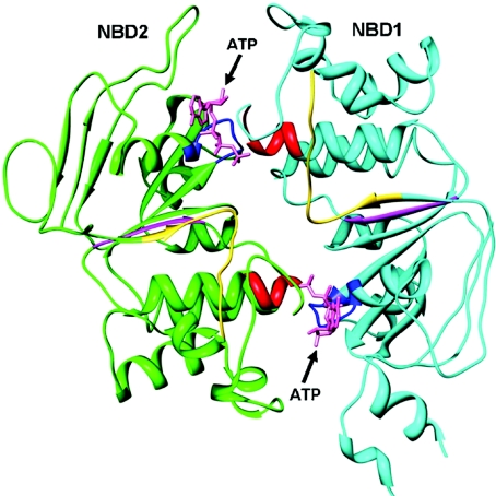
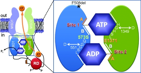
The cystic fibrosis transmembrane conductance regulator (CFTR) plays a fundamental role in fluid and electrolyte transport across epithelial tissues. Based on its structure, function and regulation, CFTR is an ATP-binding cassette (ABC) transporter. These transporters are assembled from two membrane-spanning domains (MSDs) and two nucleotide-binding domains (NBDs). In the vast majority of ABC transporters, the NBDs form a common engine that utilises the energy of ATP hydrolysis to pump a wide spectrum of substrates through diverse transmembrane pathways formed by the MSDs. By contrast, in CFTR the MSDs form a pathway for passive anion flow that is gated by cycles of ATP binding and hydrolysis by the NBDs. Here, we consider how the interaction of ATP with two ATP-binding sites, formed by the NBDs, powers conformational changes in CFTR structure to gate the channel pore. We explore how conserved sequences from both NBDs form ATP-binding sites at the interface of an NBD dimer and highlight the distinct roles that each binding site plays during the gating cycle. Knowledge of how ATP gates the CFTR Cl- channel is critical for understanding CFTR's physiological role, its malfunction in disease and the mechanism of action of small molecules that modulate CFTR channel gating.


Figures

Figure 3
Figure 3
Conformational changes of the CFTR Cl channel during channel gating The simplified model shows a CFTR Cl channel under quiescent (left) and activated (right) conditions. Communication between the NBDs and MSDs via the intracellular loops is either vertical (e.g. NBD1–MSD1) or orthogonal (e.g. NBD1–MSD2). Abbreviations: MSD, membrane-spanning domain; NBD, nucleotide-binding domain; P, phosphorylation of the RD; Pi, inorganic phosphate; PKA, cAMP-dependent protein kinase; PPase, protein phosphatase; RD, regulatory domain. In and Out denote the intra- and extracellular sides of the membrane, respectively. See text for further information. Modified, with permission, from Chen et al. (2006); ©2006 S. Karger AG, Basel.
Figure 2
Figure 2
The human CFTR NBD1–NBD2 heterodimer modelled by protein–protein docking NBD1 and NBD2, viewed from above, are represented by ribbons and coloured in cyan (NBD1) and green (NBD2). The Walker A (blue), Walker B (magenta) and LSGGQ (red) motifs are shown as well as the Q loop (yellow). ATP molecules (pink) are represented in stick mode. Reproduced, with permission, from Huang et al. (2009).
Figure 1
Figure 1
Organisation of the ATP-binding sites in CFTR The simplified model shows the molecular architecture of ATP-binding site 1 (site 1) and ATP-binding site 2 (site 2) in an open CFTR Cl channel. Each ATP binding site is formed by the Walker A and B motifs (labelled A and B, respectively) of one NBD and the LSGGQ motifs of the other NBD. Site 2 contains a canonical LSGGQ motif, whereas site 1 contains a non-canonical LSGGQ motif (LSHGH). Site 2 also contains a catalytic base (E1371) at the distal end of the Walker B motif, but this residue is absent in site 1 (S573). The location of the CF mutations G551D (site 2), G1349D (site 1) and F508del (surface of NBD1 opposing ICL4) are shown. Abbreviations: MSD, membrane-spanning domain; NBD, nucleotide-binding domain; P, phosphorylation of the RD; Pi, inorganic phosphate; RD, regulatory domain. In and Out denote the intra- and extracellular sides of the membrane, respectively. See text for further information. Modified, with permission, from Chen et al. (2006); ©2006 S. Karger AG, Basel.
Figure 4
Figure 4
2′-Deoxy-ATP potentiates strongly the single-channel activity of wild-type and G551D-CFTR A and B, representative recordings of wild-type and G551D-CFTR Cl channels in excised inside-out membrane patches in the presence of either ATP (1 mm) or 2′-deoxy-ATP (2′-dATP; 1 mm). PKA (75 nm) was continuously present in the intracellular solution, voltage was −50 mV and there was a large Cl concentration gradient across the membrane patch ([Cl]int= 147 mm; [Cl]ext= 10 mm). Dotted lines indicate where channels are closed and downward deflections of the traces correspond to channel openings. Modified, with permission, from Cai et al. (2006); ©2006 The American Society for Biochemistry and Molecular Biology.

References

    1. Aleksandrov AA, Aleksandrov L, Riordan JR. Nucleoside triphosphate pentose ring impact on CFTR gating and hydrolysis. FEBS Lett. 2002;518:183–188. - PubMed
    1. Aleksandrov AA, Aleksandrov LA, Riordan JR. CFTR (ABCC7) is a hydrolyzable-ligand-gated channel. Pflügers Arch. 2007;453:693–702. - PubMed
    1. Aleksandrov L, Aleksandrov A, Riordan JR. Mg2+-dependent ATP occlusion at the first nucleotide-binding domain (NBD1) of CFTR does not require the second (NBD2) Biochem J. 2008;416:129–136. - PubMed
    1. Anderson MP, Berger HA, Rich DP, Gregory RJ, Smith AE, Welsh MJ. Nucleoside triphosphates are required to open the CFTR chloride channel. Cell. 1991;67:775–784. - PubMed
    1. Anderson MP, Welsh MJ. Regulation by ATP and ADP of CFTR chloride channels that contain mutant nucleotide-binding domains. Science. 1992;257:1701–1704. - PubMed

Publication types

MeSH terms

Substances

LinkOut - more resources