. 2009 May 15;587(Pt 10):2353-64.
doi: 10.1113/jphysiol.2009.169656. Epub 2009 Mar 30.

Horizontal cell feedback regulates calcium currents and intracellular calcium levels in rod photoreceptors of salamander and mouse retina

Affiliations

Horizontal cell feedback regulates calcium currents and intracellular calcium levels in rod photoreceptors of salamander and mouse retina

Norbert Babai et al. J Physiol. .

Abstract

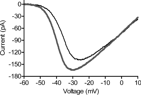
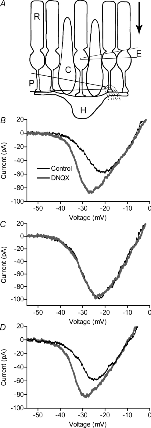
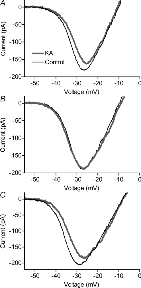
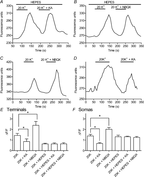
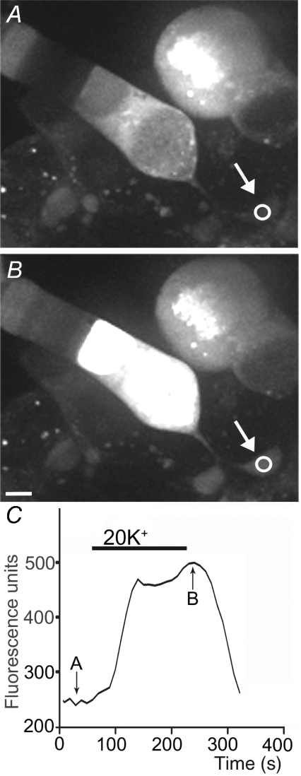
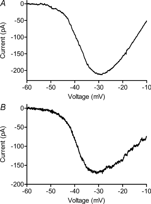
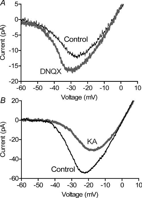
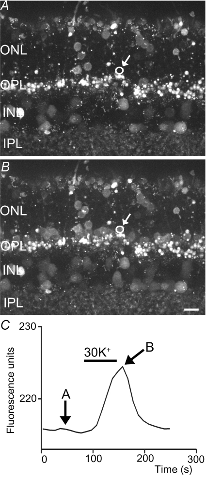
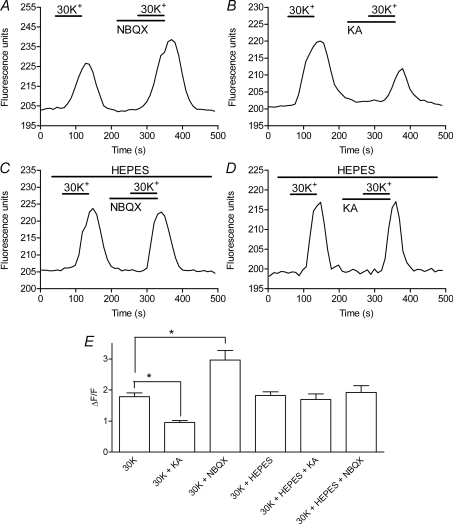
We tested whether horizontal cells (HCs) provide feedback that regulates the Ca(2+) current (I(Ca)) of rods in salamander and mouse retinas. In both species, hyperpolarizing HCs by puffing a glutamate antagonist, 6,7-dinitro-quinoxaline-2,3-dione (DNQX), onto HC processes caused a negative shift in the voltage dependence of rod I(Ca) and increased its peak amplitude. Conversely, depolarizing HCs by puffing kainic acid (KA) into the outer plexiform layer (OPL) caused a positive voltage shift and decreased rod I(Ca.) Experiments on salamander retina showed that these effects were blocked by addition of the pH buffer, Hepes. Intracellular calcium concentration ([Ca(2+)](i)) was examined in rods by confocal microscopy after loading salamander and mouse retinal slices with Fluo-4. Rods were depolarized to near the dark resting potential by bath application of high K(+) solutions. Hyperpolarizing HCs with 2,3-dihydroxy-6-nitro-7-sulphamoylbenzo[f]quinoxaline (NBQX) enhanced high K(+)-evoked Ca(2+) increases whereas depolarizing HCs with KA inhibited Ca(2+) increases. In both species these effects of NBQX and KA were blocked by addition of Hepes. Thus, like HC feedback in cones, changes in HC membrane potential modulate rod I(Ca) thereby regulating rod [Ca(2+)](i) at physiological voltages, in both mouse and salamander retinas. By countering the reduced synaptic output that accompanies hyperpolarization of rods to light, HC feedback will subtract spatially averaged luminance levels from the responses of individual rods to local changes. The finding that HC to rod feedback is present in both amphibian and mammalian species shows that this mechanism is highly conserved across vertebrate retinas.


Figures

Figure 1
Figure 1. Hyperpolarizing HCs with a saturating light flash caused a leftward shift and increase in ICa amplitude of rods recorded from flatmount salamander retina
Recording was obtained from a light-insensitive rod lacking its outer segment. In this and subsequent figures, ICa was recorded using a ramp voltage protocol (−90 to +60 mV, 0.5 mV ms−1). Thin black trace: dark. Thick grey trace: light.
Figure 2
Figure 2. Feedback effects on rod ICa of hyperpolarizing HCs with DNQX
Puffing a glutamate antagonist, DNQX (100 μm) into the OPL of salamander retina (A) caused a leftward shift and increase in peak amplitude of rod ICa (B). ICa was recorded in control conditions (thin black traces) and 525 ms after a puff (thick grey traces). As illustrated schematically in A, the puff pipette was positioned to eject DNQX into the OPL but avoid applying it to the terminal of the voltage-clamped rod. The direction of superfusate flow is indicated by the arrow. Bath application of 10 mm Hepes blocked effects of DNQX on ICa (C). Effects of DNQX recovered after washout of Hepes (D).
Figure 3
Figure 3. Feedback effects on rod ICa of depolarizing HCs with KA
Puffing a glutamate agonist, KA (1 mm), into the OPL of salamander retina caused a rightward shift and decrease in peak amplitude of rod ICa (A). Bath application of 10 mm Hepes blocked effects of KA on ICa (B). Effects of KA on ICa recovered after washout of Hepes (C). Thin black trace: control. Thick grey trace: KA.
Figure 5
Figure 5. Bath application of a glutamate antagonist NBQX or glutamate agonist KA caused [Ca2+]i changes in salamander rod terminals consistent with negative feedback effects of HC membrane potential on rod ICa
A, co-application of NBQX (3 μm) enhanced the [Ca2+]i increase in a salamander rod terminal evoked by bath application of 20 mm K+. B, co-application of KA (15 μm) inhibited the response to 20 mm K+ in a different salamander rod. Effects of NBQX and KA were both blocked by addition of 10 mm Hepes to the bathing medium (C and D). Recordings illustrated in A–D were obtained from different rod terminals in different retinal slices. E, summary of intraterminal Fluo-4 fluorescence changes evoked by high K+ relative to the basal fluorescence level measured prior to high K+ application (ΔF/F). NBQX (3 μm) significantly enhanced [Ca2+]i increases evoked by 20 mm K+ (n= 24 retinal slices, P= 0.012) whereas KA (15 μm) significantly reduced Ca2+ responses (n= 4, P= 0.041). Hepes (10 mm) blocked these effects of NBQX (n= 8, P= 0.016) and KA (n= 3, P= 0.0075). Responses to 20 K+ in the absence of these drugs were also significantly diminished by 10 mm Hepes (n= 18, P= 0.001). Data in the bar graph show changes in ΔF/F averaged from multiple retinal slices (1 data point per slice) and measurements from each slice were averaged from 1 to 8 (mean = 3.1 ± 0.23) individual rod terminals. F, similar effects were observed with high K+-evoked Fluo-4 fluorescence changes measured in rod cell bodies. NBQX (3 μm) significantly enhanced [Ca2+]i increases evoked by 20 mm K+ (n= 12 retinal slices, P= 0.011) whereas KA (15 μm) significantly reduced Ca2+ responses (n= 6, P= 0.006). Hepes (10 mm) blocked effects of NBQX (n= 7, P= 0.048) and KA (n= 9, P= 0.003). Unlike intraterminal fluorescence changes, responses to 20 mm K+ in control conditions were not diminished by 10 mm Hepes (n= 15, P= 0.80). Data in the bar graph show measurements of ΔF/F averaged from multiple retinal slices (1 data point per slice). Measurements from each slice were averaged from 1 to 5 (mean = 2.44 ± 0.16) individual rods.
Figure 4
Figure 4. Bath application of 20 mm KCl stimulated an increase in Fluo-4 fluorescence indicating an increase in [Ca2+]i in the rod synaptic terminal (arrow) and soma
Intraterminal Fluo-4 fluorescence measured in the region of interest (circle) is plotted in C. Images in A and B were obtained at the time points indicated in C. Scale bar = 5 μm.
Figure 6
Figure 6. ICa recorded from mouse (A) and salamander (B) rods were quite similar
Fitting data in the figure with a Boltzmann function adjusted for reversal potential yielded V50=−37.5 mV with slope factor =−6.76 in mouse (A) and V50=−38.5 mV with slope factor =−5.88 in salamander (B). The salamander rod lacked its outer segment.
Figure 7
Figure 7. Effects of puffing DNQX and KA on mouse rod ICa
Puffing a glutamate antagonist, DNQX (100 μm) into the OPL of mouse retina (A) caused a leftward shift and increase in peak amplitude of rod ICa. By contrast, puffing a glutamate agonist, KA (1 mm), into the OPL caused a rightward shift and decrease in peak amplitude of ICa (B). ICa was recorded in control conditions (thin black trace) and 525 ms after the puff (thick grey trace). As illustrated in Fig. 2A, the puff pipette was positioned beyond the cell so that DNQX and KA were not applied directly to the voltage-clamped rod. Data in A and B were averaged from 9 and 5 cells, respectively.
Figure 8
Figure 8. Bath application of 30 mm KCl stimulated an increase in Fluo-4 fluorescence, indicating an increase in [Ca2+]i in a mouse rod soma
Fluo-4 fluorescence measured in the region of interest (circle) is plotted in C. The images in A and B were obtained at the time points indicated in C. Scale bar = 5 μm.
Figure 9
Figure 9. Bath application of a glutamate antagonist, NBQX, or glutamate agonist, KA, caused [Ca2+]i changes in mouse rods consistent with negative feedback effects of HC membrane potential on rod ICa
A, co-application of NBQX (3 μm) enhanced the [Ca2+]i increase in a mouse rod soma evoked by bath application of 30 mm K+. B, co-application of KA (15 μm) inhibited the response to 30 mm K+. Effects of NBQX and KA were both blocked by addition of 10 mm Hepes to the bathing medium (C and D). Recordings illustrated in AD were obtained from different rods in different retinal slices. E, intracellular Fluo-4 fluorescence changes evoked by high K+ relative to the basal fluorescence level measured prior to high K+ application (ΔF/F). NBQX (3 μm) significantly enhanced [Ca2+]i increases evoked in mouse rods by 30 mm K+ (n= 18 retinal slices, P= 0.029) whereas KA (15 μm) significantly reduced Ca2+ responses (n= 15, P= 0.0001). Hepes (10 mm) blocked these effects of NBQX (n= 18, P= 0.137) and KA (n= 17; P= 0.730). Changes in ΔF/F were averaged from multiple retinal slices (1 data point per slice) and measurements from each slice were averaged from 1 to 9 (mean = 4.7 ± 0.20) individual rods.

Similar articles

Cited by

References

    1. Barnes S, Merchant V, Mahmud F. Modulation of transmission gain by protons at the photoreceptor output synapse. Proc Natl Acad Sci U S A. 1993;90:10081–10085. - PMC - PubMed
    1. Baylor DA, Fuortes MG, O’Bryan PM. Receptive fields of cones in the retina of the turtle. J Physiol. 1971;214:265–294. - PMC - PubMed
    1. Bloomfield SA, Xin D. Surround inhibition of mammalian AII amacrine cells is generated in the proximal retina. J Physiol. 2000;523:771–783. - PMC - PubMed
    1. Boycott BB, Peichl L, Wassle H. Morphological types of horizontal cell in the retina of the domestic cat. Proc R Soc Lond B Biol Sci. 1978;203:229–245. - PubMed
    1. Burkhardt DA. Responses and receptive-field organization of cones in perch retinas. J Neurophysiol. 1977;40:53–62. - PubMed

Publication types

MeSH terms

LinkOut - more resources