. 2009 Jun 19;284(25):17147-17156.
doi: 10.1074/jbc.M901031200. Epub 2009 Mar 30.

N-methyl-D-aspartate receptors mediate the phosphorylation and desensitization of muscarinic receptors in cerebellar granule neurons

Affiliations

N-methyl-D-aspartate receptors mediate the phosphorylation and desensitization of muscarinic receptors in cerebellar granule neurons

Adrian J Butcher et al. J Biol Chem. .

Abstract

Changes in synaptic strength mediated by ionotropic glutamate N-methyl-D-asparate (NMDA) receptors is generally considered to be the molecular mechanism underlying memory and learning. NMDA receptors themselves are subject to regulation through signaling pathways that are activated by G-protein-coupled receptors (GPCRs). In this study we investigate the ability of NMDA receptors to regulate the signaling of GPCRs by focusing on the G(q/11)-coupled M(3)-muscarinic receptor expressed endogenously in mouse cerebellar granule neurons. We show that NMDA receptor activation results in the phosphorylation and desensitization of M(3)-muscarinic receptors through a mechanism dependent on NMDA-mediated calcium influx and the activity of calcium-calmodulin-dependent protein kinase II. Our study reveals a complex pattern of regulation where GPCRs (M(3)-muscarinic) and NMDA receptors can feedback on each other in a process that is likely to influence the threshold value of signaling networks involved in synaptic plasticity.


Figures
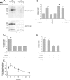
FIGURE 1.
FIGURE 1.
Activation of NMDA receptors induces phosphorylation of M3-muscarinic receptor. A, representative autoradiograph of M3-muscarinic receptors immunoprecipitated from [32P]orthophosphate labeled CG neurons prepared from wild type (WT) mice or M3-muscarinic receptor knock-out (KO) mice. The CG neurons were stimulated for 5 min with either; vehicle, methacholine (100 μm), or NMDA (100 μm). In these experiments immunoprecipitated proteins resolved by SDS-PAGE were transferred to nitrocellulose, and after exposure for phosphorylation the nitrocellulose was probed using a receptor-specific monoclonal antibody. This served as a loading control for the M3-muscarinic receptor. B, phosphorylation of the M3-muscarinic receptor in response to methacholine (100 μm), with or without atropine (10 μm), and to glutamate (100 μm) or NMDA (100 μm). C, phosphorylation of the M3-muscarinic receptor in CG neurons stimulated with the ionotropic glutamate receptor agonists NMDA, AMPA, and kainic acid (all at 100 μm). D, effect of the NMDA receptor antagonists MK-801 (5 μm, 10 min) and AP-5 (20 μm, 10 min). E, time course of M3-muscarinic receptor phosphorylation following stimulation with NMDA (100 μm). Graphs represent quantification of phosphorylation (means ± S.D.) from at least three replicates, normalized to the basal phosphorylation of the receptor in control cells with no stimulation. *, significant difference between basal and stimulated samples (p < 0.01).
FIGURE 2.
FIGURE 2.
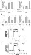
Extracellular calcium and CamKII activity is required for NMDA-mediated phosphorylation of M3-muscarinic receptors. CG neurons were [32P]orthophosphate labeled in calcium containing medium. A, cells were subsequently washed with calcium-containing (left panel) or nominally calcium-free (right panel) medium, and stimulated for 5 min with NMDA (100 μm) or methacholine (100 μm). B, phosphorylation of the M3-muscarinic receptor in response to NMDA (100 μm, 5 min) in the presence or absence of inhibitors of CamKII, KN-93 and KN-62, and the inactive analogue, KN-92 (10 μm). C and D, time course of the free intracellular calcium concentration in response to transient stimulation by methacholine or NMDA in the absence (C) or presence (D) of KN-62. The phosphorylation of M3-muscarinic receptors was quantified and normalized to the basal phosphorylation and presented as the mean ± S.D. *, significant difference between basal and stimulated samples (p < 0.01). **, significant difference between basal and stimulated samples (p < 0.05). Calcium traces correspond to the changes in free intracellular calcium in single CG neurons and represent the response of at least 20 cells in three different assays.
FIGURE 3.
FIGURE 3.
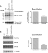
siRNA knock-down of CamKII reduces NMDA-mediated M3-muscarinic receptor phosphorylation. CG neurons on day 5 of culture were sham-transfected (NT) or transfected with scrambled siRNA duplexes or siRNA directed against CamKII-β (80 pmol). A, after a further 2 days the cells were metabolically labeled with [32P]orthophosphate and stimulated with NMDA (100 μm for 5 min). The cells were then solubilized, and M3-muscarinic receptor was immunoprecipitated. The gel was transferred to nitrocellulose and exposed to reveal the phosphorylated receptor, after which the membrane was processed by Western blot to determine equal receptor loading. B, a small sample of the lysate from each condition was retained for Western blotting for CamKII-β, GRK-2, and GRK-6. Shown are the data from a typical experiment carried out at least three times. The graphs represent the cumulative data (mean ± S.E.) of at least three experiments carried out in duplicate.
FIGURE 4.
FIGURE 4.
In vitro phosphorylation the M3-muscarinic receptor within the third intracellular loop by CamKII. A, bacterially expressed GST or GST-3iloop (containing the third intracellular loop of the mouse M3-muscarinic receptor) were used in an in vitro phosphorylation using purified CamKII (200 ng). Shown is a representative experiment where the gel is first stained with Coomassie Blue and then an autoradiograph obtained. B, diagram representation of the CamKII phospho-acceptor sites Ser322 and Ser350 as determined by LC-MS/MS (in red) and a peptide that is singularly phosphorylated at one of three sites; serines 384/385 and threonine 397 (indicated in blue). (See supplemental Figs. S2–S4 for detailed traces.)
FIGURE 5.
FIGURE 5.
NMDA-mediated phosphorylation of the M3-muscarinic receptor does not mediate receptor internalization. CG neurons were stimulated with agonists (100 μm) for the indicated times. Surface M3-muscarinic receptor expression was then determined using the hydrophilic muscarinic antagonist [3H] n-methyl scopolamine. The data represent the means ± S.D. of three experiments carried out in duplicate.
FIGURE 6.
FIGURE 6.
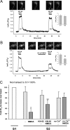
Effect of NMDA on the M3-muscarinic receptor phosphoinositide response is dependent on calcium entry and CamKII activity. CG neurons were transfected with eGFP-PHPLCδ1 and grown for 7–9 days before being imaged on an inverted epifluorescence microscope. A, continuously perfused cells were subjected to two stimulations with methacholine (100 μm). Shown is the time course of eGFP-PHPLCδ translocation from the plasma membrane to the cytoplasm in a representative single cell expressed as changes in the cytoplasmic F/F0 self-ratio (see “Experimental Procedures”). Shown also are representative fluorescent images of CG neurons stimulated with methacholine (100 μm) for 0, 3, 15, 24, and 29 min. B, same as A but including a 5-min stimulation with NMDA (100 μm) prior to the second methacholine stimulation. Shown are images of a cell at times 0, 3, 15, 18, 24, and 29 min. C, the effects of the CamKII inhibitor KN-62 (10 μm) added 10 min before NMDA or the effects of removal of extracellular calcium on the NMDA-mediated desensitization of the M3-muscarinic receptor phosphoinositide response. The data are representative traces of 12–18 cells taken from 8–10 independent experiments. The insets represent the means ± S.D. of the S1 and S2 phosphoinositide responses. *, significant difference between S1 and S2 (p < 0.01). **, significant difference between S1 and S2 (p < 0.05).

Similar articles

Cited by

References

    1. Watkins J. C., Jane D. E. (2006) Br. J. Pharmacol. 147, (Suppl. 1) S100–S108 - PMC - PubMed
    1. Malenka R. C., Nicoll R. A. (1999) Science 285, 1870–1874 - PubMed
    1. Sheng M., Kim M. J. (2002) Science 298, 776–780 - PubMed
    1. Lisman J., Schulman H., Cline H. (2002) Nat. Rev. Neurosci. 3, 175–190 - PubMed
    1. Mulkey R. M., Endo S., Shenolikar S., Malenka R. C. (1994) Nature 369, 486–488 - PubMed

Publication types

MeSH terms

Substances