. 2009 Jun;29(12):3297-306.
doi: 10.1128/MCB.00366-09. Epub 2009 Mar 30.

Phospholipase D1 regulates lymphocyte adhesion via upregulation of Rap1 at the plasma membrane

Affiliations

Phospholipase D1 regulates lymphocyte adhesion via upregulation of Rap1 at the plasma membrane

Adam Mor et al. Mol Cell Biol. 2009 Jun.

Abstract

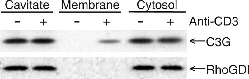
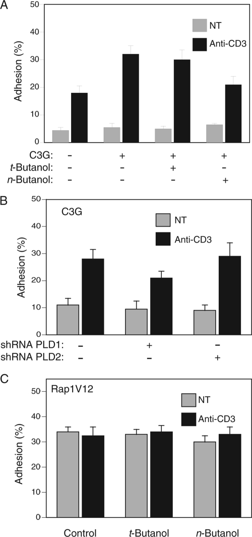
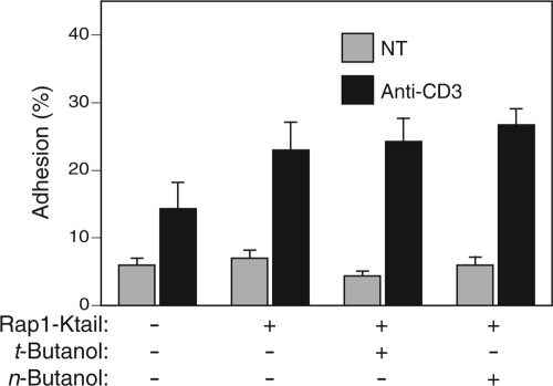
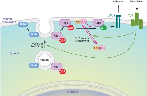
Rap1 is a small GTPase that modulates adhesion of T cells by regulating inside-out signaling through LFA-1. The bulk of Rap1 is expressed in a GDP-bound state on intracellular vesicles. Exocytosis of these vesicles delivers Rap1 to the plasma membrane, where it becomes activated. We report here that phospholipase D1 (PLD1) is expressed on the same vesicular compartment in T cells as Rap1 and is translocated to the plasma membrane along with Rap1. Moreover, PLD activity is required for both translocation and activation of Rap1. Increased T-cell adhesion in response to stimulation of the antigen receptor depended on PLD1. C3G, a Rap1 guanine nucleotide exchange factor located in the cytosol of resting cells, translocated to the plasma membranes of stimulated T cells. Our data support a model whereby PLD1 regulates Rap1 activity by controlling exocytosis of a stored, vesicular pool of Rap1 that can be activated by C3G upon delivery to the plasma membrane.


Figures

FIG. 1.
FIG. 1.
Rap1 colocalizes with PLD1 on cytoplasmic vesicles of resting cells and on the plasma membranes of activated T cells. (A) GFP-Rap1 or GFP-TC10 (vesicular GTPase control) was coexpressed with RFP-PLD1 in Jurkat T cells, which were imaged alive with a laser scanning confocal microscope. (B) GFP-Rap1 and RFP-PLD1 were expressed in COS-1 and HeLa cells and imaged live with a laser scanning confocal microscope. Colocalization is evident on both the plasma membrane and cytoplasmic vesicles. The inset shows a high-magnification view of vesicles. (C) Endogenous Rap1 and PLD1 in Jurkat T cells were analyzed for vesicle association by nitrogen cavitation, sucrose density centrifugation, and immunoblotting. Both molecules were expressed on vesicles that have the same buoyant density profile. (D) The cytosolic (S175) and membrane (P175) fractions of the postnuclear supernatant of Jurkat cell cavitation samples were analyzed by immunoprecipitation and immunobloting (Rap1 and Ras) or by immunoblotting only (Erk), as indicated. (E) The subpopulation of serum-starved Jurkat T cells expressing neither GFP-Rap1 nor mRFP-PLD1 at the plasma membrane (20% of total) was imaged before and after stimulation with an anti-CD3 antibody. Bars, 5 μm (A and E) and 10 μm (B).
FIG. 2.
FIG. 2.
Rap1 expression on the plasma membrane depends on PLD1. (A) Inhibition of PLDs with n-butanol affects the plasma membrane (PM) localization of GFP-Rap1 but not GFP-Rap1V12. (B) n-butanol inhibits anti-CD3-stimulated translocation of both GFP-Rap1 and RFP-PLD1 from cytoplasmic vesicles to the plasma membrane. (C) Endogenous PLD1 and PLD2 in HeLa cells, detected by immunoblotting before and 72 h after expression of the indicated shRNA. (D) PLD1 knockdown in Jurkat cells, shown by cytofluorimetry of cells expressing GFP-PLD1 (x axis) and the indicated shRNA, which also directs expression of RFP from an internal ribosome entry site (y axis). (E) Silencing PLD1 but not PLD2 expression with shRNA inhibits plasma membrane localization of GFP-Rap1. Images show representative cells (bars, 5 μm), and the bar graph shows means ± standard errors of the means (SEM) (n = 3) for the percentage of cells with plasma membrane expression of each fluorescent protein.
FIG. 3.
FIG. 3.
Activation of Rap1 requires PLD activity. (A) Jurkat T cells were serum starved for 2 h and treated with or without t-butanol or n-butanol for 15 min prior to stimulation with anti-CD3 antibodies for 10 min, as indicated. Cell lysates were immunoblotted for total Rap1 and GTP-Rap1 (GST-RalGDS-RBD pulldown assay). (B) Jurkat T cells expressing YFP-RalGDS-RBD, a probe for GTP-bound Rap1, and the indicated Rap1 construct were imaged live growing in serum. (C) Serum-starved Jurkat T cells expressing YFP-RalGDS-RBD and Rap1 were treated with or without t-butanol or n-butanol and imaged live before and after stimulation with anti-CD3 antibodies. Bars, 5 μm. (D) Quantification of the results shown in panel C, plotted as means± SEM (n = 4).
FIG. 4.
FIG. 4.
PLD activity is required for TCR-stimulated lymphocyte adhesion to ICAM-1. (A) Jurkat T cells transfected with the indicated Rap1 construct tagged with GFP were plated on ICAM-1 and stimulated with or without anti-CD3 antibodies, and adherent, transduced cells were measured with a fluorescent plate reader. Untransfected Jurkat cells (B) or CD4+ splenocytes from C57BL/6 mice (C) were labeled with 5-carboxyfluorescein, treated with or without t-butanol or n-butanol, and stimulated with or without anti-CD3 antibodies, and adhesion to ICAM-1 was measured as in panel A. (D) Jurkat cells labeled with 5-carboxyfluorescein and transfected with the indicated shRNA were stimulated with or without anti-CD3 antibodies, and adhesion to ICAM-1 was measured as in panel A. Results are shown as means ± SEM (n = 4), except in panel C, where the results of an experiment representative of two are shown.
FIG. 5.
FIG. 5.
TCR activation stimulates translocation of C3G from cytosol to membranes of Jurkat T cells. Serum-starved Jurkat T cells were stimulated with or without anti-CD3 antibodies and disrupted by nitrogen cavitation. The cavitated samples were separated into total membranes (P175) and cytosol (S175) and analyzed for C3G and RhoGDI by immunoblotting. Results shown are representative of three independent experiments.
FIG. 6.
FIG. 6.
C3G-dependent TCR-stimulated T-cell adhesion to ICAM-1 requires PLD1. (A) Jurkat T cells transfected with GFP-C3G or GFP vector and treated with or without t-butanol or n-butanol were assayed for adhesion to ICAM-1 as described in the legend to Fig. 5, before and after stimulation with anti-CD3 antibodies. (B) Jurkat T cells were transfected with GFP-C3G and the indicated shRNA, and the adhesion of transduced, fluorescent cells to ICAM-1 was determined. (C) Jurkat T cells transfected with GFP-Rap1V12 and treated with or without t-butanol or n-butanol were stimulated or not with anti-CD3 antibodies, and adhesion to ICAM-1 was measured. Results are shown as means ± SEM (n = 3).
FIG. 7.
FIG. 7.
Rap1 targeted directly to the plasma membrane with the Kras tail regulates T-cell adhesion in a PLD-independent fashion. Jurkat T cells were transfected with GFP vector or GFP-Rap1-Ktail, as indicated, and adhesion to ICAM-1 was measured with or without stimulation with anti-CD3 antibodies and with or without pretreatment with either t- or n-butanol. Results are shown as means ± SEM (n = 3).
FIG. 8.
FIG. 8.
Model of Rap1 upregulation and activation at the plasma membrane. In resting cells, Rap1 is stored on a pool of intracellular, endocytic vesicles in an inactive, GDP-bound state. PLD1 is expressed on the same pool of vesicles. T-cell activation leads to PLD-dependent exocytosis of these vesicles, which results in increased expression of Rap1 at the plasma membrane. TCR signaling induces concomitant translocation of C3G in a complex with an SH2/SH3 adapter, such as Crk, from the cytosol to the plasma membrane such that both the GTPase and its cognate GEF meet only at the plasma membrane, coupling upregulation to activation and leading to increased T-cell adhesion.

Similar articles

Cited by

References

    1. Amsen, D., A. Kruisbeek, J. L. Bos, and K. Reedquist. 2000. Activation of the Ras-related GTPase Rap1 by thymocyte TCR engagement and during selection. Eur. J. Immunol. 302832-2841. - PubMed
    1. Anikeeva, N., K. Somersalo, T. N. Sims, V. K. Thomas, M. L. Dustin, and Y. Sykulev. 2005. Distinct role of lymphocyte function-associated antigen-1 in mediating effective cytolytic activity by cytotoxic T lymphocytes. Proc. Natl. Acad. Sci. USA 1026437-6442. - PMC - PubMed
    1. Bergmeier, W., T. Goerge, H. W. Wang, J. R. Crittenden, A. C. Baldwin, S. M. Cifuni, D. E. Housman, A. M. Graybiel, and D. D. Wagner. 2007. Mice lacking the signaling molecule CalDAG-GEFI represent a model for leukocyte adhesion deficiency type III. J. Clin. Investig. 1171699-1707. - PMC - PubMed
    1. Bi, K., M. G. Roth, and N. T. Ktistakis. 1997. Phosphatidic acid formation by phospholipase D is required for transport from the endoplasmic reticulum to the Golgi complex. Curr. Biol. 7301-307. - PubMed
    1. Bivona, T. G., H. H. Wiener, I. M. Ahearn, J. Silletti, V. K. Chiu, and M. R. Philips. 2004. Rap1 up-regulation and activation on plasma membrane regulates T cell adhesion. J. Cell Biol. 164461-470. - PMC - PubMed

Publication types

MeSH terms

LinkOut - more resources