. 2009 Aug;23(8):2681-90.
doi: 10.1096/fj.08-128116. Epub 2009 Mar 30.

Biomarker system for studying muscle, stem cells, and cancer in vivo

Affiliations

Biomarker system for studying muscle, stem cells, and cancer in vivo

Koichi Nishijo et al. FASEB J. 2009 Aug.

Abstract

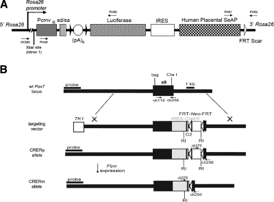
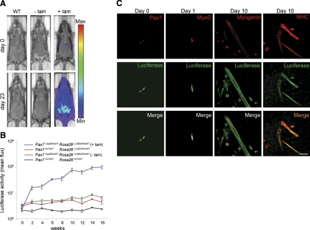
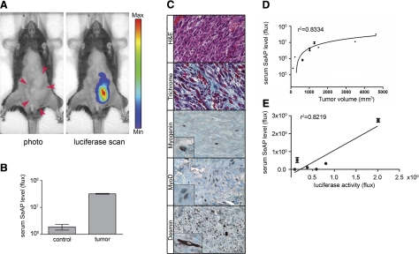
Bioluminescent reporter genes are sensitive in situ tools for following disease progression in preclinical models, albeit they are subject to scattering and absorption in deep tissues. We have generated a bicistronic Cre/LoxP reporter mouse line that pairs the expression of firefly luciferase with quantifiable expression of a human placental alkaline phosphatase that is secreted into the serum (SeAP). With the use of this dual-modality bioreporter with a novel, inducible Pax7-CreER line for tracking muscle satellite cells, we demonstrate the longitudinal kinetics of muscle stem cell turnover, accounting for a doubling of the signal from satellite cell and progeny every 3.93 wk in the transition from adolescence to early adulthood. We also show that this dual-modality bioreporter can be incorporated in preclinical cancer models, whereby SeAP activity is reflective of tumor burden. Thus, this dual bioreporter permits both spatial localization and accurate quantification of biological processes in vivo even when the tissue of interest is deep within the animal.


Figures

Figure 1.
Figure 1.
Structure of targeted mouse lines and AAVcre. A) LUSEAP dual-modality bioreporter was targeted to the Rosa26 locus at the XbaI site in intron 1. In addition to the native Rosa26 promoter, the CMV immediate-early promoter/enhancer (PcmvIE) and the SV40 late viral protein gene 16S/19S splice donor and acceptor (sd/sa) signal sites were added to strengthen reporter gene activity. To minimize leakage, a stop cassette consisting of 6 tandem copies of the SV40 viral early and late polyadenylation signal sequences [(pA)6] were inserted downstream of the promoter elements, flanked by LoxP sites (black arrowheads). Stop cassette is followed by firefly luciferase, a human IRES , and human placental secreted alkaline phosphatase. White arrowhead indicates FRT scar. B) For the Pax7-CreER allele, a targeting vector was designed for insertion of a complex cassette into the 3′ region of the Pax7 gene. At the ClaI site in exon 10, an IRES-CreER was inserted to allow bicistronic expression of Pax7 and CreER. An FRT-Neo-FRT cassette was inserted 3′ to IRES-CreER. The CRERp allele indicates the presence of the neomycin resistance gene (Neo+), whereas the CRERm allele indicated the absence of the neomycin-resistance gene (Neo) after breeding to a Flp-e deleter mouse line . RI, EcoRI restriction site.
Figure 2.
Figure 2.
Expression of reporter protein in satellite cells and their progeny using a stringent satellite cell-specific Cre allele. A) Whole-mount immunofluorescence of single myofibers explanted from Pax7CreERp/WTRosa26tm1Sor/WT mice treated with tamoxifen. Satellite cell specificity for the Pax7CreER allele is demonstrated by coexpression of the Cre reporter βGal in cells that also express the satellite cell marker Pax7 (arrowheads, top 4 panels). Some satellite cells (Pax7+) did not express βGal (arrowheads, bottom 4 panels). Scale bar = 100 μm. B) Immunohistochemistry of skeletal muscle from Pax7CreERp/WTRosa26tm1(EYFP)Cos mouse (2 wk after tamoxifen injection). Pax7-expressing cell (red) and eYFP-expressing cell (green) are colocalized beneath basement membrane. C) Immunohistochemistry for MyoD and eYFP of skeletal muscle from Pax7CreERp/WTRosa26tm1(EYFP)Cos mouse (2 wk after first tamoxifen injection). These myoblasts/satellite cell progeny (green) express MyoD (red) and localizes inside of basal lamina. Scale bar = 50 μm.
Figure 3.
Figure 3.
Application of the dual-modality bioreporter for stem cell biology using a stringent satellite cell-specific Cre allele. A) Representative example of qualitative luciferase signal from satellite cells in Pax7CreERp/WTRosa26LUSEAPm/WT mice treated with a tamoxifen pulse at 6 wk of age vs. controls. Bioluminescence is seen predominantly from the ventral (abdominal) musculature but only in tamoxifen-treated animals. Images are displayed at a minimum-maximum scale of 2 × 10⁵ to 5 × 10⁶ photons/s/cm/sr. B) Time course of quantitative bioluminescence for Pax7CreERp/WTRosa26LUSEAPm/WT mice after tamoxifen injection. Tamoxifen was intraperitoneally injected at 4 wk after birth and then luciferase signal was analyzed every 2 wk. Lines represent Pax7CreERp/WTRosa26LUSEAPm/WT (with tamoxifen; blue; n=7), Rosa26LUSEAPm/WT (green; n=4), Pax7CreERp/WTRosa26LUSEAPm/WT (without tamoxifen; red; n=5), and wild type (black; n=4), respectively. C) Immunocytochemistry of cultured myogenic progenitors from Pax7CreERp/WTRosa26LUSEAPm/WT mice. Luciferase expression was detected in Pax7-expressing satellite cells (column 1), MyoD-expressing myoblasts (column 2), myogenin-expressing immature myotubes (row 3), and MHC-expressing mature myotubes (row 4).
Figure 4.
Figure 4.
Application of dual-modality bioreporter for cancer biology. A) Abdominal tumor arising in a Pax7CreERp/WTPax3P3Fm/P3Fm Trp53F2-10/F2-10Rosa26LUSEAPm/WT 6-allele mouse allows direct comparison of luciferase and SeAP from dual-modality bioreporter. Tumor is indicated in photograph by red arrows. In luciferase scan panel, bioluminescence from the primary tumor is clearly observed with minimal background luciferase signal in the normal tissue. Image is displayed at a minimum-maximum scale of 3 × 10⁷ to 2 × 10⁸ photons/s/cm2/sr. B) Serum SeAP activity of the tumor-bearing animal (same animal as in A) is 26-fold higher than an age-matched nontumor animal with the same genotype (P<0.001). C) Histopathology confirmed the diagnosis of rhabdomyosarcoma. Hematoxylin and eosin staining shows the primary tumor. MyoD and Desmin are strongly positive, whereas myogenin is focally positive. Gomori trichrome staining indicates a collagen-rich stroma. D, E) Positive correlation was seen between serum SeAP level and tumor volumes (D) and between serum SeAP level and luciferase intensity (E) in Pax7CreERp/WTPax3P3Fm/P3FmTrp53F2-10/F2-10Rosa26LUSEAPm/WT tumor mice.

References

    1. Massoud T F, Gambhir S S. Molecular imaging in living subjects: seeing fundamental biological processes in a new light. Genes Dev. 2003;17:545–580. - PubMed
    1. Safran M, Kim W Y, Kung A L, Horner J W, DePinho R A, Kaelin W G., Jr Mouse reporter strain for noninvasive bioluminescent imaging of cells that have undergone Cre-mediated recombination. Mol Imaging. 2003;2:297–302. - PubMed
    1. Lyons S K, Meuwissen R, Krimpenfort P, Berns A. The generation of a conditional reporter that enables bioluminescence imaging of Cre/loxP-dependent tumorigenesis in mice. Cancer Res. 2003;63:7042–7046. - PubMed
    1. Nilsson E E, Westfall S D, McDonald C, Lison T, Sadler-Riggleman I, Skinner M K. An in vivo mouse reporter gene (human secreted alkaline phosphatase) model to monitor ovarian tumor growth and response to therapeutics. Cancer Chemother Pharmacol. 2002;49:93–100. - PubMed
    1. Chaudhuri T R, Cao Z, Krasnykh V N, Stargel A V, Belousova N, Partridge E E, Zinn K R. Blood-based screening and light based imaging for the early detection and monitoring of ovarian cancer xenografts. Technol Cancer Res Treat. 2003;2:171–180. - PubMed

Publication types

LinkOut - more resources