. 2009 May 20;27(15):2542-52.
doi: 10.1200/JCO.2008.19.9356. Epub 2009 Mar 30.

Edema control by cediranib, a vascular endothelial growth factor receptor-targeted kinase inhibitor, prolongs survival despite persistent brain tumor growth in mice

Affiliations

Edema control by cediranib, a vascular endothelial growth factor receptor-targeted kinase inhibitor, prolongs survival despite persistent brain tumor growth in mice

Walid S Kamoun et al. J Clin Oncol. .

Abstract

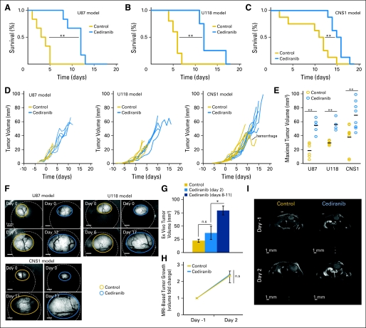
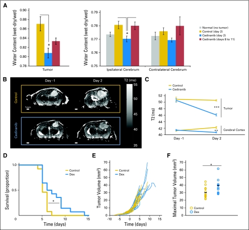
Purpose: Recent clinical trials of antivascular endothelial growth factor (VEGF) agents for glioblastoma showed promising progression-free and overall survival rates. However, available clinical imaging does not separate antitumor effects from antipermeability effects of these agents. Thus although anti-VEGF agents may decrease tumor contrast-enhancement, vascularity, and edema, the mechanisms leading to improved survival in patients remain incompletely understood. Our goal was to determine whether alleviation of edema by anti-VEGF agents alone could increase survival in mice.

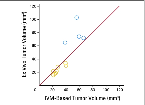
Methods: We treated mice bearing three different orthotopic models of glioblastoma with a VEGF-targeted kinase inhibitor, cediranib. Using intravital microscopy, molecular techniques, and magnetic resonance imaging (MRI), we measured survival, tumor growth, edema, vascular morphology and function, cancer cell apoptosis and proliferation, and circulating angiogenic biomarkers.

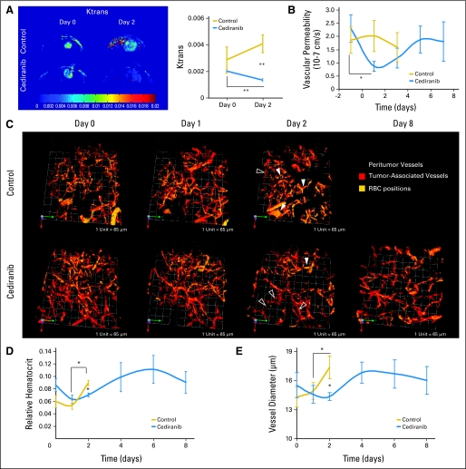
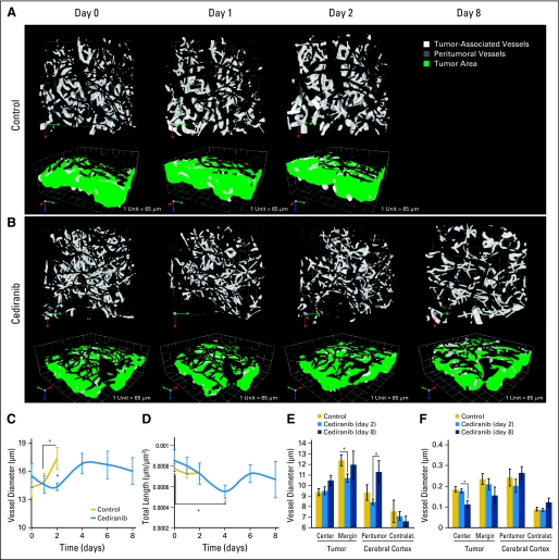
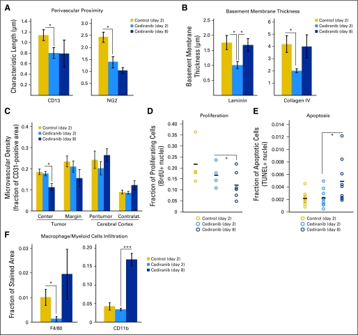
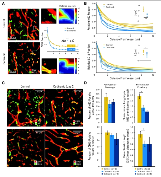
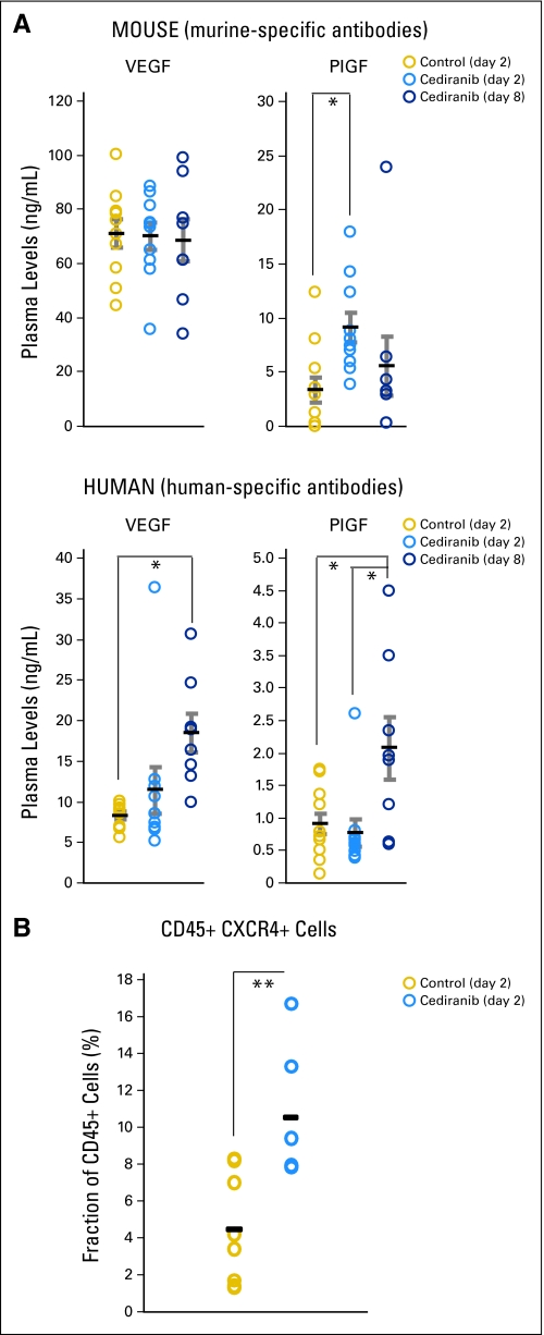
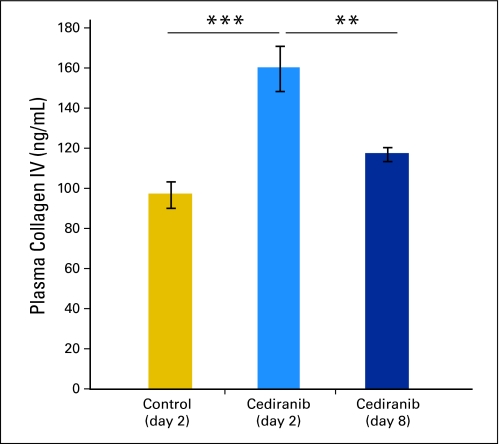
Results: We show by intravital microscopy that cediranib significantly decreased tumor vessel permeability and diameter. Moreover, cediranib treatment induced normalization of perivascular cell coverage and thinning of the basement membrane, as mirrored by an increase in plasma collagen IV. These rapid changes in tumor vascular morphology and function led to edema alleviation -- as measured by MRI and by dry/wet weight measurement of water content -- but did not affect tumor growth. By immunohistochemistry, we found a transient decrease in macrophage infiltration and significant but minor changes in tumor cell proliferation and apoptosis. Systemically, cediranib increased plasma VEGF and placenta growth factor levels, and the number of circulating CXCR4(+)CD45(+) cells. However, by controlling edema, cediranib significantly increased survival of mice in the face of persistent tumor growth.

Conclusion: Anti-VEGF agents may be able to improve survival of patients with glioblastoma, even without inhibiting tumor growth.


Conflict of interest statement

Authors' disclosures of potential conflicts of interest and author contributions are found at the end of this article.

Figures

Fig 1.
Fig 1.
Cediranib treatment leads to increased survival without effects on tumor growth. Cediranib (6 mg/kg of body weight per day) treatment leads to statistically significant survival benefit in U87 (A; **P < .001, n = 6), U118 (B; **P < .01, n = 4), and CNS1 (C; **P < .01, n = 8). (D) Single animal tumor growth curves acquired by fluorescence microscopic imaging show the lack of growth delay after cediranib treatment. (E) Maximal tumor volume (measured at end point). Cediranib-treated animals survived with statistically significantly larger U87 (**P < .001, n = 6), U118 (**P < .001, n = 4), and CNS1 (**P < .001, n = 8) tumors. (F) Representative intravital fluorescence microscopic image of U87, U118, and CNS1 at first and last time points. (G) U87 tumor volume measured ex vivo at day 2 and day 8 to 11 in control and cediranib-treated animals shows the lack of tumor growth delay with cediranib treatment at day 2 (P > .05, n = 5) (H) Cediranib treatment does not affect U87 tumor growth assessed by magnetic resonance imaging (volumes measured on T2 weighted stacks 0.15 mm XY resolution × 0.5 mm Z resolution; n = 3) (I) Representative T2-weighted single brain slices of cediranib- and control-treated animals bearing U87 tumors.
Fig 2.
Fig 2.
Cediranib decreases glioblastoma-induced edema, and corticosteroid-mediated edema control increases survival without affecting tumor growth. (A) Water content measured by dry/wet weight ratio in the tumor and ipsilateral and contralateral cerebrum. Cediranib significantly decreased tumor and ipsilateral water content (*P < .05, n = 5). (B) Representative T2 maps of single brain slices used to evaluate edema (T2 correlates with water content). (C) Quantification of T2 in the tumor and the cerebral cortex. Cediranib significantly decreased T2 signal in the tumor (***P < .001) and the cerebral cortex (**P < .01). (D) Dexamethasone (Dex) (10 mg/kg each day) treatment leads to a statistically significant survival benefit (*P < .05, n = 14). (E) Individual tumor growth curves acquired by fluorescence microscopic imaging showing the lack of growth delay after Dex treatment. (F) Maximal tumor volume (measured at end point). Dex-treated animals survived with significantly larger tumors (*P < .05, n = 14).
Fig 3.
Fig 3.
Cediranib treatment normalizes glioma vessel morphology and function. (A) Cediranib treatment (6 mg/kg each day) leads to a statistically significant decrease in Ktrans (arbitrary units, **P < .05, n = 4). (B) Cediranib significantly decreases vascular permeability measured by intravital microscopy (*P < .05, n = 12). (C) Representative multiphoton laser scanning microscopy (MPLSM) three-dimensional reconstruction of the tumor (red) and peritumor vessels (gray) superimposed with RBC positions acquired through analysis of injected fluorescently labeled RBC. Control animals have predominantly hemoconcentrated vessels (closed arrows). Cediranib-treated animals have predominantly low hematocrit vessels (open arrows). (D) Mean and SE of tumor vessel relative hematocrit measured by MPLSM. Cediranib significantly decreases relative hematocrit (*P < .05, n = 4). (E) Cediranib transiently but significantly decreases vessel diameter measured by MPLSM (*P < .05 n = 4).
Fig 4.
Fig 4.
Cediranib decreases vessel diameter at early time points and decreases vessel density at later time points. (A, B) Representative multiphoton laser scanning microscopic (MPLSM) three-dimensional reconstruction of the tumor (white) and peritumor vessels (gray) showing the effects of (A) control versus (B) cediranib treatment on vessel diameter. (C) Cediranib transiently but significantly decreases vessel diameter measured by MPLSM (*P < .05, n = 4). (D) Cediranib significantly decreases vessel density (length) measured by MPLSM (*P < .05, n = 4). (E) Cediranib decreases vessel diameter at the tumor margin at day 2 (measured by immunostaining for CD31 endothelial staining; *P < .05, n = 9). (F) Cediranib decreases microvascular density at the tumor center at day 8 (measured by immunostaining for CD31; *P < .05, n = 9).
Fig 5.
Fig 5.
Cediranib treatment decreases macrophage infiltration and normal vessel wall structure; at day 8, microvascular density is decreased, tumor cell proliferation is decreased, and apoptosis is increased. (A) Cediranib decreases the characteristic length describing the distance between perivascular cells and the vessel wall (measured by immunostaining for CD13, *P < .05, n = 5; and NG2, *P < .05, n = 9). (B) Cediranib transiently but significantly decreases basement membrane thickness at day 2 (measured by immunostaining for laminin and collagen IV; *P < .05, n = 5). (C) Cediranib decreases microvascular density at the tumor center at day 8 (measured by immunostaining for CD31 endothelial staining; *P < .05, n = 9). (D) A total of 1 mg/mouse of 5-bromo-2-deoxyuridine (BrdU) was injected intraperitoneally 24 hours before the end point. The fraction of nuclei with incorporated BrdU is quantified and plotted for each animal. Cediranib significantly decreased proliferation only at day 8 (*P < .05, n = 4). (E) Terminal deoxynucleotidyl transferase–mediated (TUNEL) staining was used to assess apoptosis of tumor cells. The fraction of TUNEL-positive nuclei was plotted for each animal. Cediranib significantly increased apoptosis at day 8 (*P < .05, n = 9). (F) Cendiranib transiently but significantly decreases macrophages infiltration at day 2 (measured by immunostaining for F4/80; *P < .05, n = 5). Myeloid cells infiltration is significantly increased at day 8 (measured by immunostaining for CD11b; **P < .0001, n = 5).
Fig 6.
Fig 6.
Quantification of pericyte coverage and proximity. (A) Representative immunohistochemistry fields of CD31- (endothelial marker) and NG2- (pericyte marker) stained tumor vessels. The fraction of NG2-positive pixels was analyzed at various distances from the vessel wall (1 μm to 10 μm), and the data were fit to an exponential function, yielding the quantity L, the characteristic extension of stain from the wall. (B) The fraction of NG2- or CD13- (pericyte marker) positive pixels was analyzed at various distances from the vessel wall. The average signal for each animal (based on five areas) is plotted, showing a difference between cediranib-treated and control animals. (C) Representative field of CD31- (endothelial marker) and NG2- (top panels) or CD13- (bottom panels) stained tumor vessels with control or cediranib treatment at day 2. (D) Cediranib significantly decreased pericyte distance from endothelial cells without changing the extent of perivascular cell coverage (NG2, P < .05, n = 9; CD13, P < .05, n = 5).
Fig A1.
Fig A1.
Validation of intravital measurements (IVM) of tumor volume. To validate the accuracy of the tumor volume estimation, we compared tumor volume measurements based on fluorescence microscopy imaging to actual tumor weights measured ex vivo. This comparison was performed for U87 tumors and showed that the green fluorescence protein–based estimations are highly accurate for small tumor volumes (approximately 20 μL reached at day 2, yellow circles, ie, when the comparisons with untreated tumors were performed), but showed a tendency to underestimate the tumor volume for larger tumor sizes (reached at day 8 after cediranib treatment, blue circles).
Fig A2.
Fig A2.
Systemic effects of cediranib treatment. Levels of human- and mouse-derived vascular endothelial growth factor (VEGF) and placenta growth factor (PlGF) in plasma of mice with orthotopic U87 gliomas, plotted for control treatment (day 2) and cediranib treatment (at days 2 and 8). Cediranib significantly increased the plasma levels of both mouse and human (tumor-derived) PlGF (*P < .05) and human VEGF (*P < .05), as well as the number of circulating CXCR4+CD45+ cells (**P < .01).
Fig A3.
Fig A3.
Changes in plasma collagen IV levels mirror thinning of the basement membrane. We hypothesized that the thinning of the vascular basement membrane owing to vascular normalization would increase the level of circulating collagen IV. Indeed, cediranib significantly increased the plasma levels of collagen IV at day 2 (***P < .001 cediranib day 2 v control). Collagen IV levels were decreased at day 8 (**P < .01 cediranib day 2 v cediranib day 8).

References

    1. Batchelor TT, Sorensen AG, di Tomaso E, et al. AZD2171, a pan-VEGF receptor tyrosine kinase inhibitor, normalizes tumor vasculature and alleviates edema in glioblastoma patients. Cancer Cell. 2007;11:83–95. - PMC - PubMed
    1. Cloughesy TF. A phase II, randomized, non-comparative clinical trial of the effect of bevacizumab (BV) alone or in combination with irinotecan (CPT) on 6-month progression free survival (PFS6) in recurrent, treatment-refractory glioblastoma (GBM) J Clin Oncol. 2008;26(suppl):91s. abstr 2010b.
    1. Pope WB, Lai A, Nghiemphu P, et al. MRI in patients with high-grade gliomas treated with bevacizumab and chemotherapy. Neurology. 2006;66:1258–1260. - PubMed
    1. Vredenburgh JJ, Desjardins A, Herndon JE, 2nd, et al. Phase II trial of bevacizumab and irinotecan in recurrent malignant glioma. Clin Cancer Res. 2007;13:1253–1259. - PubMed
    1. Norden AD, Young GS, Setayesh K, et al. Bevacizumab for recurrent malignant gliomas: Efficacy, toxicity, and patterns of recurrence. Neurology. 2008;70:779–787. - PubMed

Publication types

MeSH terms