

A multi-marker assay to distinguish malignant melanomas from benign nevi
- PMID: 19332774
- PMCID: PMC2662963
- DOI: 10.1073/pnas.0901185106
A multi-marker assay to distinguish malignant melanomas from benign nevi
Abstract
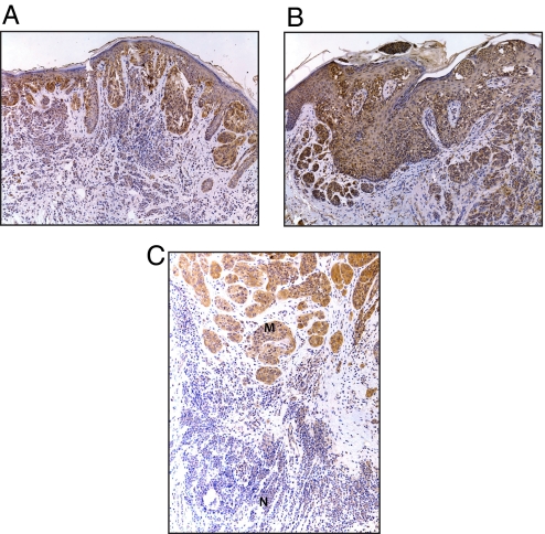
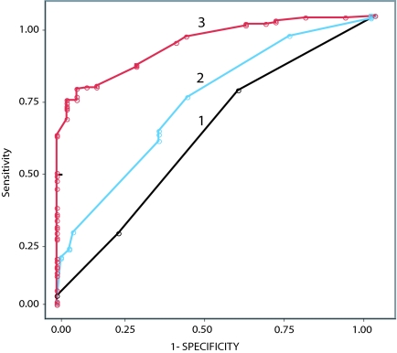
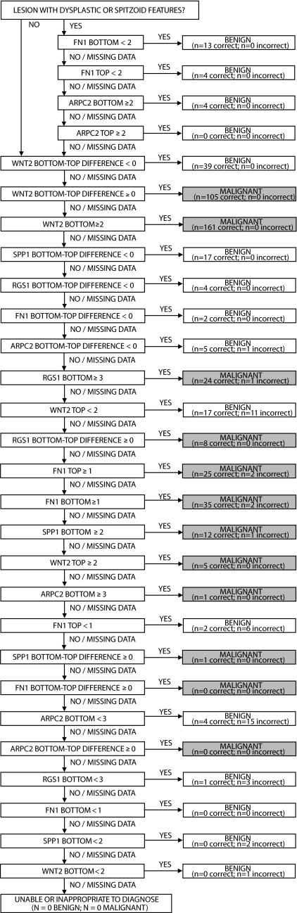
The histopathological diagnosis of melanoma can be challenging. No currently used molecular markers accurately distinguish between nevus and melanoma. Recent transcriptome analyses have shown the differential expression of several genes in melanoma progression. Here, we describe a multi-marker diagnostic assay using 5 markers (ARPC2, FN1, RGS1, SPP1, and WNT2) overexpressed in melanomas. Immunohistochemical marker expression was analyzed in 693 melanocytic neoplasms comprising a training set (tissue microarray of 534 melanomas and nevi), and 4 independent validation sets: tissue sections of melanoma arising in a nevus; dysplastic nevi; Spitz nevi; and misdiagnosed melanocytic neoplasms. Both intensity and pattern of expression were scored for each marker. Based on the differential expression of these 5 markers between nevi and melanomas in the training set, a diagnostic algorithm was obtained. Using this algorithm, the lesions in the validation sets were diagnosed as nevus or melanoma, and the results were compared with the known histological diagnoses. Both the intensity and pattern of expression of each marker were significantly different in melanomas compared to nevi. The diagnostic algorithm exploiting these differences achieved a specificity of 95% and a sensitivity of 91% in the training set. In the validation sets, the multi-marker assay correctly diagnosed a high percentage of melanomas arising in a nevus, Spitz nevi, dysplastic nevi, and misdiagnosed lesions. The multi-marker assay described here can aid in the diagnosis of melanoma.
Conflict of interest statement
Conflict of interest statement: M.K.-S. has stock in Melanoma Diagnostics, Inc. J.R.M. has ownership interest in MDMS.
Figures
References
-
- Jemal A, et al. Cancer statistics, 2008. CA Cancer J Clin. 2008;58:71–96. - PubMed
-
- Crowson AN, Magro CM, Mihm MC., Jr . The Melanocytic Proliferations. New York: Wiley-Liss; 2001. Chapter 10.
-
- Piepkorn MW, et al. A multiobserver, population-based analysis of histologic dysplasia in melanocytic nevi. J Am Acad Dermatol. 1994;30:707–714. - PubMed
-
- Farmer ER, Gonin R, Hanna MP. Discordance in the histopathologic diagnosis of melanoma and melanocytic nevi between expert pathologists. Hum Pathol. 1996;27:528–531. - PubMed
-
- Corona R, et al. Interobserver variability on the histopathologic diagnosis of cutaneous melanoma and other pigmented skin lesions. J Clin Oncol. 1996;14:1218–1223. - PubMed
Publication types
MeSH terms
Substances
Grants and funding
LinkOut - more resources
Full Text Sources
Other Literature Sources
Medical
Research Materials
Miscellaneous
