. 2009 Apr 14;106(15):6303-8.
doi: 10.1073/pnas.0809422106. Epub 2009 Mar 30.

PD-1 expression by macrophages plays a pathologic role in altering microbial clearance and the innate inflammatory response to sepsis

Affiliations

PD-1 expression by macrophages plays a pathologic role in altering microbial clearance and the innate inflammatory response to sepsis

Xin Huang et al. Proc Natl Acad Sci U S A. .

Abstract

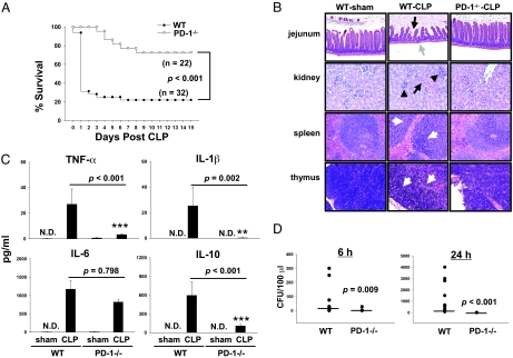
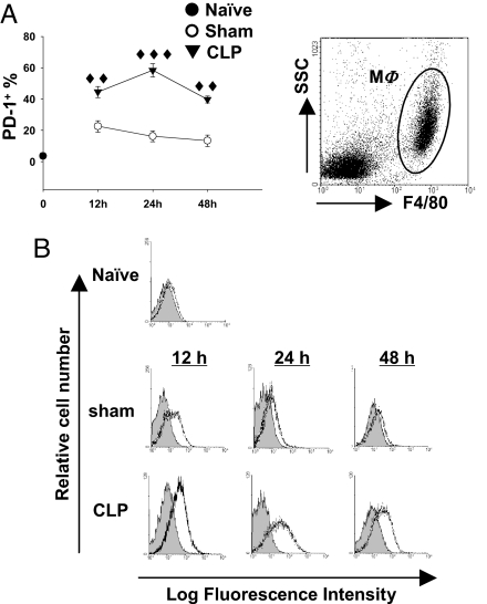
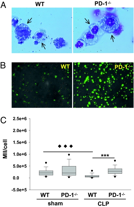
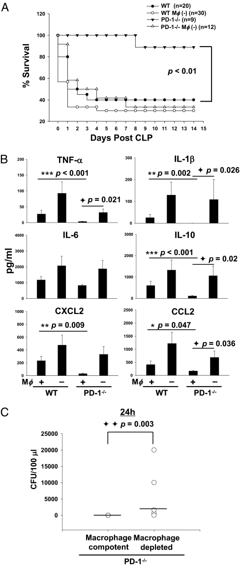
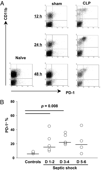
Sepsis, a leading cause of death worldwide, involves concomitant expression of an overzealous inflammatory response and inefficient bacterial clearance. Macrophage function is pivotal to the development of these two aspects during sepsis; however, the mechanisms underlying these changes remain unclear. Here we report that the PD-1:PD-L pathway appears to be a determining factor of the outcome of sepsis, regulating the delicate balance between effectiveness and damage by the antimicrobial immune response. To this end we observed that PD-1(-/-) mice were markedly protected from the lethality of sepsis, accompanied by a decreased bacterial burden and suppressed inflammatory cytokine response. To the extent that this is a macrophage-specific aspect of the effects of PD-1, we found the following: first, peritoneal macrophages expressed significantly higher levels of PD-1 during sepsis, which was associated with their development of cellular dysfunction; second, when peritoneal macrophages were depleted (using clodronate liposomes) from PD-1(-/-) mice, the animals' bactericidal capacity was lowered, their inflammatory cytokine levels were elevated, and protection from septic lethality was diminished; and third, blood monocytes from both septic mice and patients with septic shock shared markedly increased PD-1 levels. Together, these data suggest that PD-1 may not only be a dysfunctional marker/effector of macrophages/monocytes, but may also be a potential therapeutic target for designing measures to modulate the innate immune response, thereby preventing the detrimental effects of sepsis.


Conflict of interest statement

The authors declare no conflict of interest.

Figures

Fig. 1.
Fig. 1.
PD-1 deficiency protects mice from sepsis-induced lethality and displayed reduced organ damage, a less severe cytokine storm, and improved bacterial clearance. (A) PD-1−/− mice were resistant to CLP-treated as compared with WT mice. WT (n = 32) and PD-1−/− (n = 22) mice were subjected to CLP, and survival was monitored for 15 days. P < 0.001 by log-rank test. (B) PD-1−/− mice showed less histological evidence of tissue destruction than did WT mice during severe sepsis. PD-1−/− mice and WT were subjected to CLP surgery or sham control surgery. Mice were euthanized 40 hours after surgery. Tissues were stainined with hematoxylin and eosin. Original magnification, ×200 for jejunum and ×400 for kidney, spleen, and thymus. (Jejunum) Black arrow and gray arrow point to examples of villus shortening and mucosal wall thinning in WT septic mice, respectively; PD-1−/− septic mice do not show these pathologic conditions. (Kidney) Black arrow and arrowheads indicate acute tubular necrosis and congested glomeruli, respectively, in a kidney from a WT septic mouse. Kidney from PD-1−/− septic mice showed rare tubular necrosis and much less congestion. (Spleen and thymus) White arrows illustrate areas of kayrorrhectic, pycnotic, apoptotic cell bodies, which are absent in PD-1−/− septic mice. Data are representative of four to eight mice per group. (C) PD-1−/− mice showed significantly lower systemic levels of inflammatory cytokines at 24 hours post-CLP than did WT mice. Graphs depict data (mean ± SEM of 4–8 sham and 11–13 CLP) pooled from three independent experiments. N.D., not detectable. **P < 0.01 and *** P < 0.001, CLP-treated PD-1−/− mice vs. CLP-treated WT mice by Mann-Whitney test. (D) PD-1−/− mice had markedly reduced bacterial burden after CLP. Levels of aerobic bacteria were expressed as CFU per 100 μl. The graphs depict data pooled from two to three independent studies showing similar results (n = 9–11). Horizontal bar indicates median for each group. P value by Mann-Whitney test.
Fig. 2.
Fig. 2.
Expression of PD-1 in macrophages is augmented by sepsis. (A) The percentage of PD-1+ macrophage increased during sepsis. PD-1 expression on peritoneal macrophages (gated as F4/80+) was determined by flow cytometry (n = 5–12 for sham-treated mice, n = 7–17 for CLP-treated mice pooled from two to three experiments). ♦♦, P < 0.01, ♦♦♦, P < 0.001 by Mann-Whitney test, CLP-treated WT mice vs. sham-treated WT mice. (B) Representative histograms of PD-1 expression on peritoneal macrophages. Peritoneal macrophages were gated as F4/80+ peritoneal leukocytes (Upper). PD-1 expression (black lines) was overlayed on isotype control (gray filled; Lower).
Fig. 3.
Fig. 3.
Sepsis-induced macrophage dysfunction is associated with the expression of PD-1. (A) Septic mouse macrophages from WT mice showed significant impairment in bacterial clearance, whereas septic PD-1−/− macrophages did not. Peritoneal leukocytes were collected at 24 hours post-CLP, spun on slides, and stained with Giemsa staining solution. WT macrophages (arrows) displayed an exhausted phenotype in which bacteria accumulated within the cell plasma, whereas PD-1−/− septic mouse macrophages (arrows) exhibited an activated phenotype. Representative images from three independent experiments show similar results. Original magnification, × 200. (B and C) Phagocytosis assay of macrophages. (B) Representative images of septic peritoneal macrophages from WT or PD-1−/− mice after feeding with Fluorescein-conjugated E. coli. Light microscope images were overlayed with fluorescence images. (C) Quantitative analysis of phagocytosis. Approximately 150 cells from sham group (n = 2 of WT or PD-1−/− mice) or 300 cells from CLP group (n = 3 of WT or PD-1−/− mice) were calculated by transforming individual cells from image into mean integrated intensity (MII) of fluorescence per cell. Data are representative of a total of two experiments. Outliers are shown at 5th/95th percentiles. ◆◆◆ P < 0.001, CLP-treated WT mice vs. sham treated WT mice.*** P < 0.001, CLP-treated PD-1−/− mice vs. CLP-treated WT mice, by Mann-Whitney test.
Fig. 4.
Fig. 4.
Macrophage is an important mediator of the protective effects of PD-1 gene deficiency in septic mice. (A) Macrophages are pivotal in maintaining the protective effect of PD-1 gene deficiency in mice from the lethality of sepsis. WT and PD-1−/− mice were treated with clodronate liposomes or PBS (200 μl/mouse, i.p.) 48 hours before being subjected to CLP. The survival rate was followed for 15 days after surgery. Graph represents survival data pooled from two experiments. (B) Peritoneal macrophages are critical for regulating the cytokine response seen in PD-1−/− mice during sepsis. Concentrations of cytokines in plasma (sham, n = 4; CLP, n = 12–16) were measured by ELISA. * P < 0.05, ** P < 0.01, and *** P < 0.001, CLP-treated PD-1−/− mice vs. CLP-treated WT mice; ◆ P < 0.05, macrophage depleted PD-1−/− mice vs. macrophage intact PD-1−/− mice by Mann-Whitney test. Graphs depict data (mean ± SEM) pooled from two to three independent experiments. (C) Macrophages play a role in the improved bacterial clearance seen in PD-1−/− mice during sepsis. At 24 hours after CLP, macrophage-competent mice and macrophage-depleted mice were bled, spread on TSA plates, and incubated for 24–48 hours at 37 °C. Levels of bacteria were expressed as CFU per 100 μl. Horizontal bar indicates median for each group. (n = 5–9) P value by Mann-Whitney test.
Fig. 5.
Fig. 5.
PD-1 may be a tolerance marker for circulating monocytes in sepsis. (A) PD-1 expression on circulating mouse blood monocytes was up-regulated after CLP. Representative data of PD-1 expression on mouse blood monocytes from naïve mice or mice subjected to sham or CLP surgery at indicated time points. (B) PD-1 expression on circulating monocytes was higher in patients with septic shock than in healthy volunteers. Horizontal bar indicates the median for each group. P value by Mann-Whitney test.

Comment in

References

    1. Angus DC, et al. Epidemiology of severe sepsis in the United States: Analysis of incidence, outcome, and associated costs of care. Crit Care Med. 2001;29:1303–1310. - PubMed
    1. Martin GS, Mannino DM, Eaton S, Moss M. The epidemiology of sepsis in the United States from 1979 through 2000. N Engl J Med. 2003;348:1546–1554. - PubMed
    1. Cohen J. The immunopathogenesis of sepsis. Nature. 2002;420:885–891. - PubMed
    1. Wheeler AP, Bernard GR. Treating patients with severe sepsis. N Engl J Med. 1999;340:207–214. - PubMed
    1. Annane D, Bellissant E, Cavaillon JM. Septic shock. Lancet. 2005;365:63–78. - PubMed

Publication types

MeSH terms